 Movie
Movie Controller


+ Open data

- Basic information
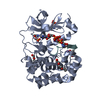
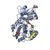
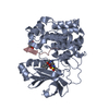
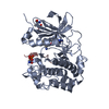
| Entry | Database: PDB / ID: 6buu | |||||||||
|---|---|---|---|---|---|---|---|---|---|---|
| Title | Crystal structure of AKT1 (aa 144-480) with a bisubstrate | |||||||||
 Components Components |
| |||||||||
 Keywords Keywords | TRANSFERASE / AKT1 / RAC-alpha serine/theronine-protein kinase / kinase / phosphorylated tail / semi-synthesis | |||||||||
| Function / homology |  Function and homology information Function and homology informationpositive regulation of endodeoxyribonuclease activity / regulation of tRNA methylation / negative regulation of protein maturation / negative regulation of fatty acid beta-oxidation / positive regulation of protein localization to endoplasmic reticulum / regulation of glycogen biosynthetic process / negative regulation of lymphocyte migration / cellular response to decreased oxygen levels / cellular response to rapamycin / negative regulation of protein localization to lysosome ...positive regulation of endodeoxyribonuclease activity / regulation of tRNA methylation / negative regulation of protein maturation / negative regulation of fatty acid beta-oxidation / positive regulation of protein localization to endoplasmic reticulum / regulation of glycogen biosynthetic process / negative regulation of lymphocyte migration / cellular response to decreased oxygen levels / cellular response to rapamycin / negative regulation of protein localization to lysosome / maternal placenta development / maintenance of protein location in mitochondrion / mammalian oogenesis stage / AKT-mediated inactivation of FOXO1A / negative regulation of long-chain fatty acid import across plasma membrane / Negative regulation of the PI3K/AKT network / regulation of type B pancreatic cell development / positive regulation of anaphase-promoting complex-dependent catabolic process / : / potassium channel activator activity / sperm glycocalyx / AKT phosphorylates targets in the nucleus / activation-induced cell death of T cells / mammary gland epithelial cell differentiation / negative regulation of glycogen (starch) synthase activity / neuron projection organization / regulation of microtubule anchoring at centrosome / negative regulation of mesenchymal stem cell differentiation / negative regulation of type B pancreatic cell development / cellular response to oxidised low-density lipoprotein particle stimulus / negative regulation of cilium assembly / negative regulation of hydrogen peroxide-induced neuron intrinsic apoptotic signaling pathway / Butyrate Response Factor 1 (BRF1) binds and destabilizes mRNA / fibroblast migration / superior temporal gyrus development / positive regulation of protein localization to cilium / negative regulation of glycogen biosynthetic process / positive regulation of TORC2 signaling / positive regulation of glucose metabolic process / perinuclear theca / negative regulation of TORC2 signaling / RUNX2 regulates genes involved in cell migration / cellular response to peptide / beta-arrestin-dependent dopamine receptor signaling pathway / negative regulation of dopaminergic neuron differentiation / positive regulation of organ growth / positive regulation of protein localization to centrosome / maintenance of cell polarity / regulation of protein export from nucleus / interleukin-18-mediated signaling pathway / positive regulation of sodium ion transport / positive regulation of cilium assembly / MTOR signalling / response to growth factor / heart valve development / CRMPs in Sema3A signaling / tau-protein kinase / cell migration involved in sprouting angiogenesis / beta-catenin destruction complex / peripheral nervous system myelin maintenance / response to fluid shear stress / cellular response to granulocyte macrophage colony-stimulating factor stimulus / negative regulation of leukocyte cell-cell adhesion / RAB GEFs exchange GTP for GDP on RABs / APC truncation mutants have impaired AXIN binding / AXIN missense mutants destabilize the destruction complex / Truncations of AMER1 destabilize the destruction complex / glycogen biosynthetic process / positive regulation of mitochondrial outer membrane permeabilization involved in apoptotic signaling pathway / phosphatidylinositol-3,4-bisphosphate binding / positive regulation of protein localization to cell surface / cellular response to interleukin-3 / Maturation of nucleoprotein / Beta-catenin phosphorylation cascade / Signaling by GSK3beta mutants / CTNNB1 S33 mutants aren't phosphorylated / CTNNB1 S37 mutants aren't phosphorylated / CTNNB1 S45 mutants aren't phosphorylated / CTNNB1 T41 mutants aren't phosphorylated / phosphorylation / complement receptor mediated signaling pathway / negative regulation of TOR signaling / regulation of long-term synaptic potentiation / sphingosine-1-phosphate receptor signaling pathway / Wnt signalosome / regulation of microtubule-based process / response to growth hormone / AKT phosphorylates targets in the cytosol / anoikis / regulation of postsynapse organization / Disassembly of the destruction complex and recruitment of AXIN to the membrane / positive regulation of fibroblast migration / labyrinthine layer blood vessel development / positive regulation of protein binding / regulation of axon extension / negative regulation of calcineurin-NFAT signaling cascade / negative regulation of protein localization to nucleus / execution phase of apoptosis / Maturation of nucleoprotein / regulation of myelination Similarity search - Function | |||||||||
| Biological species |  Homo sapiens (human) Homo sapiens (human) | |||||||||
| Method |  X-RAY DIFFRACTION / FOURIER SYNTHESIS / Resolution: 2.4 Å X-RAY DIFFRACTION / FOURIER SYNTHESIS / Resolution: 2.4 Å | |||||||||
| Model details | Mn+bisubstrate | |||||||||
 Authors Authors | Chu, N. / Gabelli, S.B. / Cole, P.A. | |||||||||
| Funding support |  United States, 2items United States, 2items
| |||||||||
 Citation Citation | Journal: Cell / Year: 2018 Title: Akt Kinase Activation Mechanisms Revealed Using Protein Semisynthesis. Authors: Chu, N. / Salguero, A.L. / Liu, A.Z. / Chen, Z. / Dempsey, D.R. / Ficarro, S.B. / Alexander, W.M. / Marto, J.A. / Li, Y. / Amzel, L.M. / Gabelli, S.B. / Cole, P.A. | |||||||||
| History |
|
- Structure visualization
| Structure viewer | Molecule: MolmilJmol/JSmol |
|---|
- Downloads & links
-Download
| PDBx/mmCIF format | 6buu.cif.gz | 159.1 KB | Display | PDBx/mmCIF format |
|---|---|---|---|---|
| PDB format | pdb6buu.ent.gz | 122.4 KB | Display | PDB format |
| PDBx/mmJSON format | 6buu.json.gz | Tree view | PDBx/mmJSON format | |
| Others |  Other downloads Other downloads |
-Validation report
| Arichive directory | https://data.pdbj.org/pub/pdb/validation_reports/bu/6buuftp://data.pdbj.org/pub/pdb/validation_reports/bu/6buu | HTTPS FTP |
|---|
-Related structure data
| Related structure data |  6npzC  4ekkS S: Starting model for refinement C: citing same article ( |
|---|---|
| Similar structure data |


-Links
 PDBj
PDBj


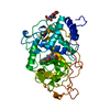
- Assembly
| Deposited unit | 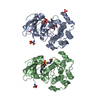
| ||||||||
|---|---|---|---|---|---|---|---|---|---|
| 1 | 
| ||||||||
| 2 | 
| ||||||||
| Unit cell |
|
-Components
| #1: Protein | Mass: 39238.488 Da / Num. of mol.: 2 Source method: isolated from a genetically manipulated source Source: (gene. exp.) Homo sapiens (human) / Gene: AKT1, PKB, RAC / Plasmid: pfastbacProduction host:  Spodoptera aff. frugiperda 2 RZ-2014 (butterflies/moths) Spodoptera aff. frugiperda 2 RZ-2014 (butterflies/moths)Strain (production host): SF9 References: UniProt: P31749, non-specific serine/threonine protein kinase #2: Protein/peptide | Mass: 1685.502 Da / Num. of mol.: 2 / Source method: obtained synthetically / Source: (synth.) Homo sapiens (human) / References: UniProt: P49841*PLUS#3: Chemical | ChemComp-SO4 / |   Mass: 96.063 Da / Num. of mol.: 1 / Source method: obtained synthetically / Formula: SO4 Mass: 96.063 Da / Num. of mol.: 1 / Source method: obtained synthetically / Formula: SO4#4: Chemical |   Mass: 54.938 Da / Num. of mol.: 2 / Source method: obtained synthetically / Formula: Mn Mass: 54.938 Da / Num. of mol.: 2 / Source method: obtained synthetically / Formula: Mn#5: Water | ChemComp-HOH / |  Mass: 18.015 Da / Num. of mol.: 238 / Source method: isolated from a natural source / Formula: H2O Mass: 18.015 Da / Num. of mol.: 238 / Source method: isolated from a natural source / Formula: H2OHas protein modification | Y | |
|---|
-Experimental details
-Experiment
| Experiment | Method: X-RAY DIFFRACTION / Number of used crystals: 1 |
|---|
- Sample preparation
| Crystal | Density Matthews: 2.68 Å3/Da / Density % sol: 54.18 % / Mosaicity: 1.039 ° / Mosaicity esd: 0.014 ° |
|---|---|
| Crystal grow | Temperature: 277 K / Method: vapor diffusion / pH: 7.5 Details: 20% PEG 3000, 0.1M Hepes HCL, 0.2M Ammonium sulfate |
-Data collection
| Diffraction | Mean temperature: 100 K | |||||||||||||||||||||||||||||||||||||||||||||||||||||||||||||||||||||||||||||||||||||||||||||||||||||||||||||||||||||||||||||||||||||||||||||||||||||||||||||||||||||||||||||||||||||||||||||
|---|---|---|---|---|---|---|---|---|---|---|---|---|---|---|---|---|---|---|---|---|---|---|---|---|---|---|---|---|---|---|---|---|---|---|---|---|---|---|---|---|---|---|---|---|---|---|---|---|---|---|---|---|---|---|---|---|---|---|---|---|---|---|---|---|---|---|---|---|---|---|---|---|---|---|---|---|---|---|---|---|---|---|---|---|---|---|---|---|---|---|---|---|---|---|---|---|---|---|---|---|---|---|---|---|---|---|---|---|---|---|---|---|---|---|---|---|---|---|---|---|---|---|---|---|---|---|---|---|---|---|---|---|---|---|---|---|---|---|---|---|---|---|---|---|---|---|---|---|---|---|---|---|---|---|---|---|---|---|---|---|---|---|---|---|---|---|---|---|---|---|---|---|---|---|---|---|---|---|---|---|---|---|---|---|---|---|---|---|---|---|
| Diffraction source | Source: ROTATING ANODE / Type: RIGAKU FR-E+ DW / Wavelength: 1.54187 Å | |||||||||||||||||||||||||||||||||||||||||||||||||||||||||||||||||||||||||||||||||||||||||||||||||||||||||||||||||||||||||||||||||||||||||||||||||||||||||||||||||||||||||||||||||||||||||||||
| Detector | Type: DECTRIS PILATUS3 R 200K-A / Detector: PIXEL / Date: Oct 4, 2016 | |||||||||||||||||||||||||||||||||||||||||||||||||||||||||||||||||||||||||||||||||||||||||||||||||||||||||||||||||||||||||||||||||||||||||||||||||||||||||||||||||||||||||||||||||||||||||||||
| Radiation | Protocol: SINGLE WAVELENGTH / Monochromatic (M) / Laue (L): M / Scattering type: x-ray | |||||||||||||||||||||||||||||||||||||||||||||||||||||||||||||||||||||||||||||||||||||||||||||||||||||||||||||||||||||||||||||||||||||||||||||||||||||||||||||||||||||||||||||||||||||||||||||
| Radiation wavelength | Wavelength: 1.54187 Å / Relative weight: 1 | |||||||||||||||||||||||||||||||||||||||||||||||||||||||||||||||||||||||||||||||||||||||||||||||||||||||||||||||||||||||||||||||||||||||||||||||||||||||||||||||||||||||||||||||||||||||||||||
| Reflection | Resolution: 2.4→50 Å / Num. obs: 31906 / % possible obs: 94.4 % / Redundancy: 3 % / Rmerge(I) obs: 0.07 / Rpim(I) all: 0.046 / Rrim(I) all: 0.084 / Χ2: 2.995 / Net I/σ(I): 17.5 / Num. measured all: 96011 | |||||||||||||||||||||||||||||||||||||||||||||||||||||||||||||||||||||||||||||||||||||||||||||||||||||||||||||||||||||||||||||||||||||||||||||||||||||||||||||||||||||||||||||||||||||||||||||
| Reflection shell | Diffraction-ID: 1
|
- Processing
| Software |
| ||||||||||||||||||||||||||||||||||||||||||||||||||||||||||||
|---|---|---|---|---|---|---|---|---|---|---|---|---|---|---|---|---|---|---|---|---|---|---|---|---|---|---|---|---|---|---|---|---|---|---|---|---|---|---|---|---|---|---|---|---|---|---|---|---|---|---|---|---|---|---|---|---|---|---|---|---|---|
| Refinement | Method to determine structure: FOURIER SYNTHESIS Starting model: 4EKK Resolution: 2.4→47 Å / Cor.coef. Fo:Fc: 0.943 / Cor.coef. Fo:Fc free: 0.924 / WRfactor Rfree: 0.2268 / WRfactor Rwork: 0.1818 / FOM work R set: 0.839 / SU B: 6.834 / SU ML: 0.162 / SU R Cruickshank DPI: 0.5097 / SU Rfree: 0.2665 / Cross valid method: THROUGHOUT / σ(F): 0 / ESU R: 0.51 / ESU R Free: 0.267 / Stereochemistry target values: MAXIMUM LIKELIHOOD Details: HYDROGENS HAVE BEEN ADDED IN THE RIDING POSITIONS U VALUES : REFINED INDIVIDUALLY
| ||||||||||||||||||||||||||||||||||||||||||||||||||||||||||||
| Solvent computation | Ion probe radii: 0.8 Å / Shrinkage radii: 0.8 Å / VDW probe radii: 1.2 Å / Solvent model: MASK | ||||||||||||||||||||||||||||||||||||||||||||||||||||||||||||
| Displacement parameters | Biso max: 150.93 Å2 / Biso mean: 37.814 Å2 / Biso min: 8.88 Å2
| ||||||||||||||||||||||||||||||||||||||||||||||||||||||||||||
| Refinement step | Cycle: final / Resolution: 2.4→47 Å
| ||||||||||||||||||||||||||||||||||||||||||||||||||||||||||||
| Refine LS restraints |
| ||||||||||||||||||||||||||||||||||||||||||||||||||||||||||||
| LS refinement shell | Resolution: 2.398→2.46 Å / Rfactor Rfree error: 0 / Total num. of bins used: 20
|
