 Movie
Movie Controller


+ Open data

- Basic information
| Entry | Database: PDB / ID: 1h8f | ||||||
|---|---|---|---|---|---|---|---|
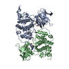
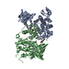
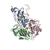
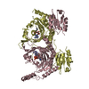
| Title | Glycogen Synthase Kinase 3 beta. | ||||||
 Components Components | GLYCOGEN SYNTHASE KINASE-3 BETA | ||||||
 Keywords Keywords | KINASE / INSULIN PATHWAY | ||||||
| Function / homology |  Function and homology information Function and homology informationnegative regulation of glycogen (starch) synthase activity / neuron projection organization / regulation of microtubule anchoring at centrosome / negative regulation of mesenchymal stem cell differentiation / negative regulation of type B pancreatic cell development / superior temporal gyrus development / positive regulation of protein localization to cilium / negative regulation of glycogen biosynthetic process / negative regulation of TORC2 signaling / beta-arrestin-dependent dopamine receptor signaling pathway ...negative regulation of glycogen (starch) synthase activity / neuron projection organization / regulation of microtubule anchoring at centrosome / negative regulation of mesenchymal stem cell differentiation / negative regulation of type B pancreatic cell development / superior temporal gyrus development / positive regulation of protein localization to cilium / negative regulation of glycogen biosynthetic process / negative regulation of TORC2 signaling / beta-arrestin-dependent dopamine receptor signaling pathway / negative regulation of dopaminergic neuron differentiation / positive regulation of protein localization to centrosome / maintenance of cell polarity / regulation of protein export from nucleus / positive regulation of cilium assembly / heart valve development / CRMPs in Sema3A signaling / tau-protein kinase / beta-catenin destruction complex / APC truncation mutants have impaired AXIN binding / AXIN missense mutants destabilize the destruction complex / Truncations of AMER1 destabilize the destruction complex / positive regulation of mitochondrial outer membrane permeabilization involved in apoptotic signaling pathway / cellular response to interleukin-3 / Maturation of nucleoprotein / Beta-catenin phosphorylation cascade / Signaling by GSK3beta mutants / CTNNB1 S33 mutants aren't phosphorylated / CTNNB1 S37 mutants aren't phosphorylated / CTNNB1 S45 mutants aren't phosphorylated / CTNNB1 T41 mutants aren't phosphorylated / negative regulation of TOR signaling / regulation of long-term synaptic potentiation / Wnt signalosome / regulation of microtubule-based process / AKT phosphorylates targets in the cytosol / Disassembly of the destruction complex and recruitment of AXIN to the membrane / positive regulation of protein binding / regulation of axon extension / negative regulation of calcineurin-NFAT signaling cascade / negative regulation of protein localization to nucleus / Maturation of nucleoprotein / negative regulation of epithelial to mesenchymal transition / tau-protein kinase activity / positive regulation of cell-matrix adhesion / glycogen metabolic process / ER overload response / regulation of axonogenesis / regulation of dendrite morphogenesis / regulation of neuron projection development / establishment of cell polarity / Constitutive Signaling by AKT1 E17K in Cancer / protein kinase A catalytic subunit binding / dynactin binding / epithelial to mesenchymal transition / canonical Wnt signaling pathway / Regulation of HSF1-mediated heat shock response / negative regulation of osteoblast differentiation / NF-kappaB binding / negative regulation of extrinsic apoptotic signaling pathway via death domain receptors / extrinsic apoptotic signaling pathway / negative regulation of protein-containing complex assembly / regulation of cellular response to heat / cellular response to retinoic acid / extrinsic apoptotic signaling pathway in absence of ligand / positive regulation of type I interferon production / Transcriptional and post-translational regulation of MITF-M expression and activity / positive regulation of autophagy / response to endoplasmic reticulum stress / positive regulation of protein export from nucleus / presynaptic modulation of chemical synaptic transmission / negative regulation of cell migration / excitatory postsynaptic potential / positive regulation of protein ubiquitination / peptidyl-serine phosphorylation / hippocampus development / regulation of microtubule cytoskeleton organization / positive regulation of cell differentiation / mitochondrion organization / Ubiquitin-dependent degradation of Cyclin D / negative regulation of canonical Wnt signaling pathway / circadian rhythm / positive regulation of protein-containing complex assembly / regulation of circadian rhythm / GSK3B and BTRC:CUL1-mediated-degradation of NFE2L2 / beta-catenin binding / B-WICH complex positively regulates rRNA expression / Degradation of GLI2 by the proteasome / GLI3 is processed to GLI3R by the proteasome / tau protein binding / Degradation of beta-catenin by the destruction complex / cellular response to amyloid-beta / Wnt signaling pathway / neuron projection development / kinase activity / positive regulation of protein catabolic process / p53 binding / Regulation of RUNX2 expression and activity / insulin receptor signaling pathway / protein autophosphorylation Similarity search - Function | ||||||
| Biological species |  HOMO SAPIENS (human) HOMO SAPIENS (human) | ||||||
| Method |  X-RAY DIFFRACTION / SYNCHROTRON / MOLECULAR REPLACEMENT / Resolution: 2.8 Å X-RAY DIFFRACTION / SYNCHROTRON / MOLECULAR REPLACEMENT / Resolution: 2.8 Å | ||||||
 Authors Authors | Dajani, R. / Pearl, L.H. / Roe, S.M. | ||||||
 Citation Citation | Journal: Cell(Cambridge,Mass.) / Year: 2001 Title: Crystal Structure of Glycogen Synthase Kinase 3Beta . Structural Basis for Phosphate-Primed Substrate Specificity and Autoinhibition Authors: Dajani, R. / Fraser, E. / Roe, S.M. / Young, N. / Good, V. / Dale, T.C. / Pearl, L.H. | ||||||
| History |
| ||||||
| Remark 700 | SHEET DETERMINATION METHOD: DSSP THE SHEETS PRESENTED AS "AA" AND "BA" ON SHEET RECORDS BELOW ARE ... SHEET DETERMINATION METHOD: DSSP THE SHEETS PRESENTED AS "AA" AND "BA" ON SHEET RECORDS BELOW ARE ACTUALLY A 6-STRANDED BARREL THIS IS REPRESENTED BY A 7-STRANDED SHEET IN WHICH THE FIRST AND LAST STRANDS ARE IDENTICAL. |
- Structure visualization
| Structure viewer | Molecule: MolmilJmol/JSmol |
|---|
- Downloads & links
-Download
| PDBx/mmCIF format | 1h8f.cif.gz | 153.2 KB | Display | PDBx/mmCIF format |
|---|---|---|---|---|
| PDB format | pdb1h8f.ent.gz | 120.7 KB | Display | PDB format |
| PDBx/mmJSON format | 1h8f.json.gz | Tree view | PDBx/mmJSON format | |
| Others |  Other downloads Other downloads |
-Validation report
| Arichive directory | https://data.pdbj.org/pub/pdb/validation_reports/h8/1h8fftp://data.pdbj.org/pub/pdb/validation_reports/h8/1h8f | HTTPS FTP |
|---|
-Related structure data
| Related structure data |  1pmeS S: Starting model for refinement |
|---|---|
| Similar structure data |

-Links
 PDBj
PDBj


- Assembly
| Deposited unit | 
| ||||||||
|---|---|---|---|---|---|---|---|---|---|
| 1 |
| ||||||||
| Unit cell |
|
-Components
| #1: Protein | Mass: 39799.746 Da / Num. of mol.: 2 Source method: isolated from a genetically manipulated source Source: (gene. exp.) HOMO SAPIENS (human) / Plasmid: PFASTBAC HTA / Cell line (production host): Sf9 / Production host:   Spodoptera frugiperda (fall armyworm) / References: UniProt: P49841, EC: 2.7.1.37 Spodoptera frugiperda (fall armyworm) / References: UniProt: P49841, EC: 2.7.1.37#2: Chemical |   Mass: 238.305 Da / Num. of mol.: 2 / Source method: obtained synthetically / Formula: C8H18N2O4S / Comment: pH buffer*YM Mass: 238.305 Da / Num. of mol.: 2 / Source method: obtained synthetically / Formula: C8H18N2O4S / Comment: pH buffer*YM#3: Water | ChemComp-HOH / |  Mass: 18.015 Da / Num. of mol.: 161 / Source method: isolated from a natural source / Formula: H2O Mass: 18.015 Da / Num. of mol.: 161 / Source method: isolated from a natural source / Formula: H2OSequence details | HIS 350 IN SWISS-PROT SHOULD BE LEU | |
|---|
-Experimental details
-Experiment
| Experiment | Method: X-RAY DIFFRACTION / Number of used crystals: 1 |
|---|
- Sample preparation
| Crystal | Density Matthews: 3.2 Å3/Da / Density % sol: 61 % | ||||||||||||||||||||||||||||||||||||||||||||||||||||||||
|---|---|---|---|---|---|---|---|---|---|---|---|---|---|---|---|---|---|---|---|---|---|---|---|---|---|---|---|---|---|---|---|---|---|---|---|---|---|---|---|---|---|---|---|---|---|---|---|---|---|---|---|---|---|---|---|---|---|
| Crystal grow | Method: vapor diffusion, hanging drop / pH: 7.5 Details: CRYSTAL WERE GROWN BY THE HANGING DROP METHOD. 1UL OF PROTEIN SOLUTION (4MG/ML IN 20MM HEPES-NAOH, 500MM NACL, 2MM MGCL2, 1MM DTT, PH 7.2) WAS MIXED WITH 1UL PRECIPITANT (6% PEG8000, 100MM TRIS-HCL, PH 7.5) | ||||||||||||||||||||||||||||||||||||||||||||||||||||||||
| Crystal grow | *PLUS pH: 7.2 / Method: vapor diffusion, hanging drop | ||||||||||||||||||||||||||||||||||||||||||||||||||||||||
| Components of the solutions | *PLUS
|
-Data collection
| Diffraction | Mean temperature: 100 K |
|---|---|
| Diffraction source | Source: SYNCHROTRON / Site: SRS  / Beamline: PX14.2 / Wavelength: 1.488 / Beamline: PX14.2 / Wavelength: 1.488 |
| Detector | Type: ADSC CCD / Detector: CCD / Date: Sep 15, 2000 |
| Radiation | Protocol: SINGLE WAVELENGTH / Monochromatic (M) / Laue (L): M / Scattering type: x-ray |
| Radiation wavelength | Wavelength: 1.488 Å / Relative weight: 1 |
| Reflection | Resolution: 2.75→35 Å / Num. obs: 34002 / % possible obs: 99.9 % / Observed criterion σ(I): 0 / Redundancy: 5.6 % / Biso Wilson estimate: 30.7 Å2 / Rsym value: 0.074 / Net I/σ(I): 8.4 |
| Reflection shell | Resolution: 2.75→2.9 Å / Redundancy: 5.6 % / Mean I/σ(I) obs: 2.3 / Rsym value: 0.323 / % possible all: 100 |
| Reflection | *PLUS Num. obs: 33985 / Rmerge(I) obs: 0.074 |
| Reflection shell | *PLUS % possible obs: 99.9 % / Num. unique obs: 4883 / Rmerge(I) obs: 0.323 |
- Processing
| Software |
| ||||||||||||||||||||||||||||||||||||||||||||||||||||||||||||
|---|---|---|---|---|---|---|---|---|---|---|---|---|---|---|---|---|---|---|---|---|---|---|---|---|---|---|---|---|---|---|---|---|---|---|---|---|---|---|---|---|---|---|---|---|---|---|---|---|---|---|---|---|---|---|---|---|---|---|---|---|---|
| Refinement | Method to determine structure: MOLECULAR REPLACEMENT Starting model: PDB ENTRY 1PME Resolution: 2.8→35.12 Å / Rfactor Rfree error: 0.006 / Data cutoff high absF: 1640575.05 / Isotropic thermal model: RESTRAINED / Cross valid method: THROUGHOUT / σ(F): 0
| ||||||||||||||||||||||||||||||||||||||||||||||||||||||||||||
| Solvent computation | Solvent model: FLAT MODEL / Bsol: 36.7 Å2 / ksol: 0.34558 e/Å3 | ||||||||||||||||||||||||||||||||||||||||||||||||||||||||||||
| Displacement parameters | Biso mean: 58.5 Å2
| ||||||||||||||||||||||||||||||||||||||||||||||||||||||||||||
| Refine analyze |
| ||||||||||||||||||||||||||||||||||||||||||||||||||||||||||||
| Refinement step | Cycle: LAST / Resolution: 2.8→35.12 Å
| ||||||||||||||||||||||||||||||||||||||||||||||||||||||||||||
| Refine LS restraints |
| ||||||||||||||||||||||||||||||||||||||||||||||||||||||||||||
| LS refinement shell | Resolution: 2.8→2.98 Å / Rfactor Rfree error: 0.022 / Total num. of bins used: 6
| ||||||||||||||||||||||||||||||||||||||||||||||||||||||||||||
| Xplor file |
| ||||||||||||||||||||||||||||||||||||||||||||||||||||||||||||
| Software | *PLUS Name: CNS / Version: 1 / Classification: refinement | ||||||||||||||||||||||||||||||||||||||||||||||||||||||||||||
| Refinement | *PLUS Lowest resolution: 35 Å / Rfactor obs: 0.22 / Rfactor Rfree: 0.254 / Rfactor Rwork: 0.22 | ||||||||||||||||||||||||||||||||||||||||||||||||||||||||||||
| Solvent computation | *PLUS | ||||||||||||||||||||||||||||||||||||||||||||||||||||||||||||
| Displacement parameters | *PLUS | ||||||||||||||||||||||||||||||||||||||||||||||||||||||||||||
| Refine LS restraints | *PLUS
| ||||||||||||||||||||||||||||||||||||||||||||||||||||||||||||
| LS refinement shell | *PLUS Rfactor Rfree: 0.35 |
