 Movie
Movie Controller

[English] 日本語
 Yorodumi
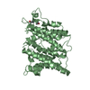
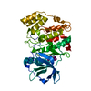
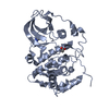
- PDB-3pup: Structure of Glycogen Synthase Kinase 3 beta (GSK3B) in complex w... -
+ Open data

- Basic information
| Entry | Database: PDB / ID: 3pup | ||||||
|---|---|---|---|---|---|---|---|
| Title | Structure of Glycogen Synthase Kinase 3 beta (GSK3B) in complex with a ruthenium octasporine ligand (OS1) | ||||||
 Components Components | Glycogen synthase kinase-3 beta | ||||||
 Keywords Keywords | TRANSFERASE / WNT SIGNALING PATHWAY / SERINE/THREONINE-PROTEIN KINASE / RUTHENIUM GLYCOGEN SYNTHASE KINASE PICOMOLAR / NUCLEOTIDE-BINDING / KINASE / ATP-BINDING / PHOSPHOPROTEIN | ||||||
| Function / homology |  Function and homology information Function and homology informationnegative regulation of glycogen (starch) synthase activity / neuron projection organization / regulation of microtubule anchoring at centrosome / negative regulation of mesenchymal stem cell differentiation / negative regulation of type B pancreatic cell development / superior temporal gyrus development / positive regulation of protein localization to cilium / negative regulation of glycogen biosynthetic process / negative regulation of TORC2 signaling / beta-arrestin-dependent dopamine receptor signaling pathway ...negative regulation of glycogen (starch) synthase activity / neuron projection organization / regulation of microtubule anchoring at centrosome / negative regulation of mesenchymal stem cell differentiation / negative regulation of type B pancreatic cell development / superior temporal gyrus development / positive regulation of protein localization to cilium / negative regulation of glycogen biosynthetic process / negative regulation of TORC2 signaling / beta-arrestin-dependent dopamine receptor signaling pathway / negative regulation of dopaminergic neuron differentiation / positive regulation of protein localization to centrosome / maintenance of cell polarity / regulation of protein export from nucleus / positive regulation of cilium assembly / heart valve development / CRMPs in Sema3A signaling / tau-protein kinase / beta-catenin destruction complex / APC truncation mutants have impaired AXIN binding / AXIN missense mutants destabilize the destruction complex / Truncations of AMER1 destabilize the destruction complex / positive regulation of mitochondrial outer membrane permeabilization involved in apoptotic signaling pathway / cellular response to interleukin-3 / Maturation of nucleoprotein / Beta-catenin phosphorylation cascade / Signaling by GSK3beta mutants / CTNNB1 S33 mutants aren't phosphorylated / CTNNB1 S37 mutants aren't phosphorylated / CTNNB1 S45 mutants aren't phosphorylated / CTNNB1 T41 mutants aren't phosphorylated / negative regulation of TOR signaling / regulation of long-term synaptic potentiation / Wnt signalosome / regulation of microtubule-based process / AKT phosphorylates targets in the cytosol / Disassembly of the destruction complex and recruitment of AXIN to the membrane / positive regulation of protein binding / regulation of axon extension / negative regulation of calcineurin-NFAT signaling cascade / negative regulation of protein localization to nucleus / Maturation of nucleoprotein / negative regulation of epithelial to mesenchymal transition / tau-protein kinase activity / positive regulation of cell-matrix adhesion / glycogen metabolic process / ER overload response / regulation of axonogenesis / regulation of dendrite morphogenesis / regulation of neuron projection development / establishment of cell polarity / Constitutive Signaling by AKT1 E17K in Cancer / protein kinase A catalytic subunit binding / dynactin binding / epithelial to mesenchymal transition / canonical Wnt signaling pathway / Regulation of HSF1-mediated heat shock response / negative regulation of osteoblast differentiation / NF-kappaB binding / negative regulation of extrinsic apoptotic signaling pathway via death domain receptors / extrinsic apoptotic signaling pathway / negative regulation of protein-containing complex assembly / regulation of cellular response to heat / cellular response to retinoic acid / extrinsic apoptotic signaling pathway in absence of ligand / positive regulation of type I interferon production / Transcriptional and post-translational regulation of MITF-M expression and activity / positive regulation of autophagy / response to endoplasmic reticulum stress / positive regulation of protein export from nucleus / presynaptic modulation of chemical synaptic transmission / negative regulation of cell migration / excitatory postsynaptic potential / positive regulation of protein ubiquitination / peptidyl-serine phosphorylation / hippocampus development / regulation of microtubule cytoskeleton organization / positive regulation of cell differentiation / mitochondrion organization / Ubiquitin-dependent degradation of Cyclin D / negative regulation of canonical Wnt signaling pathway / circadian rhythm / positive regulation of protein-containing complex assembly / regulation of circadian rhythm / GSK3B and BTRC:CUL1-mediated-degradation of NFE2L2 / beta-catenin binding / B-WICH complex positively regulates rRNA expression / Degradation of GLI2 by the proteasome / GLI3 is processed to GLI3R by the proteasome / tau protein binding / Degradation of beta-catenin by the destruction complex / cellular response to amyloid-beta / Wnt signaling pathway / neuron projection development / kinase activity / positive regulation of protein catabolic process / p53 binding / Regulation of RUNX2 expression and activity / insulin receptor signaling pathway / protein autophosphorylation Similarity search - Function | ||||||
| Biological species |  Homo sapiens (human) Homo sapiens (human) | ||||||
| Method |  X-RAY DIFFRACTION / SYNCHROTRON / MOLECULAR REPLACEMENT / molecular replacement / Resolution: 2.99 Å X-RAY DIFFRACTION / SYNCHROTRON / MOLECULAR REPLACEMENT / molecular replacement / Resolution: 2.99 Å | ||||||
 Authors Authors | Filippakopoulos, P. / Kraling, K. / Essen, L.O. / Meggers, E. / Knapp, S. | ||||||
 Citation Citation | Journal: J.Am.Chem.Soc. / Year: 2011 Title: Structurally sophisticated octahedral metal complexes as highly selective protein kinase inhibitors. Authors: Feng, L. / Geisselbrecht, Y. / Blanck, S. / Wilbuer, A. / Atilla-Gokcumen, G.E. / Filippakopoulos, P. / Kraling, K. / Celik, M.A. / Harms, K. / Maksimoska, J. / Marmorstein, R. / Frenking, G. ...Authors: Feng, L. / Geisselbrecht, Y. / Blanck, S. / Wilbuer, A. / Atilla-Gokcumen, G.E. / Filippakopoulos, P. / Kraling, K. / Celik, M.A. / Harms, K. / Maksimoska, J. / Marmorstein, R. / Frenking, G. / Knapp, S. / Essen, L.O. / Meggers, E. #1: Journal: J.Am.Chem.Soc. / Year: 2008Title: Targeting large kinase active site with rigid, bulky octahedral ruthenium complexes. Authors: Maksimoska, J. / Feng, L. / Harms, K. / Yi, C. / Kissil, J. / Marmorstein, R. / Meggers, E. #2: Journal: Chembiochem / Year: 2008Title: Extremely tight binding of a ruthenium complex to glycogen synthase kinase 3. Authors: Atilla-Gokcumen, G.E. / Pagano, N. / Streu, C. / Maksimoska, J. / Filippakopoulos, P. / Knapp, S. / Meggers, E. | ||||||
| History |
|
- Structure visualization
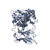
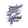
| Structure viewer | Molecule: MolmilJmol/JSmol |
|---|
- Downloads & links
-Download
| PDBx/mmCIF format | 3pup.cif.gz | 283.7 KB | Display | PDBx/mmCIF format |
|---|---|---|---|---|
| PDB format | pdb3pup.ent.gz | 227.9 KB | Display | PDB format |
| PDBx/mmJSON format | 3pup.json.gz | Tree view | PDBx/mmJSON format | |
| Others |  Other downloads Other downloads |
-Validation report
| Arichive directory | https://data.pdbj.org/pub/pdb/validation_reports/pu/3pupftp://data.pdbj.org/pub/pdb/validation_reports/pu/3pup | HTTPS FTP |
|---|
-Related structure data
| Related structure data |  2yakC  2jldS S: Starting model for refinement C: citing same article ( |
|---|---|
| Similar structure data |


-Links
 PDBj
PDBj


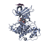
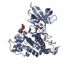
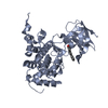
- Assembly
| Deposited unit | 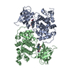
| |||||||||||||||||||||||||||||||||||||||||||||||||||||||||||||||||||||||||||||||||||||||||||||||||||
|---|---|---|---|---|---|---|---|---|---|---|---|---|---|---|---|---|---|---|---|---|---|---|---|---|---|---|---|---|---|---|---|---|---|---|---|---|---|---|---|---|---|---|---|---|---|---|---|---|---|---|---|---|---|---|---|---|---|---|---|---|---|---|---|---|---|---|---|---|---|---|---|---|---|---|---|---|---|---|---|---|---|---|---|---|---|---|---|---|---|---|---|---|---|---|---|---|---|---|---|---|
| 1 | 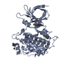
| |||||||||||||||||||||||||||||||||||||||||||||||||||||||||||||||||||||||||||||||||||||||||||||||||||
| 2 | 
| |||||||||||||||||||||||||||||||||||||||||||||||||||||||||||||||||||||||||||||||||||||||||||||||||||
| 3 |
| |||||||||||||||||||||||||||||||||||||||||||||||||||||||||||||||||||||||||||||||||||||||||||||||||||
| Unit cell |
| |||||||||||||||||||||||||||||||||||||||||||||||||||||||||||||||||||||||||||||||||||||||||||||||||||
| Noncrystallographic symmetry (NCS) | NCS domain:
NCS domain segments: Ens-ID: 1 / Refine code: 1
| |||||||||||||||||||||||||||||||||||||||||||||||||||||||||||||||||||||||||||||||||||||||||||||||||||
| Details | Monomer |
-Components
| #1: Protein | Mass: 46801.215 Da / Num. of mol.: 2 Source method: isolated from a genetically manipulated source Source: (gene. exp.) Homo sapiens (human) / Gene: GSK3B / Plasmid: pET151 / Production host:  #2: Chemical |   Mass: 572.945 Da / Num. of mol.: 2 / Source method: obtained synthetically / Formula: C24H16ClN6O3Ru Mass: 572.945 Da / Num. of mol.: 2 / Source method: obtained synthetically / Formula: C24H16ClN6O3Ru |
|---|
-Experimental details
-Experiment
| Experiment | Method: X-RAY DIFFRACTION / Number of used crystals: 1 |
|---|
- Sample preparation
| Crystal | Density Matthews: 3.34 Å3/Da / Density % sol: 63.19 % |
|---|---|
| Crystal grow | Temperature: 277 K / Method: vapor diffusion, hanging drop / pH: 7.4 Details: 100 mM Tris pH 7.4 20 % PEG 6000, VAPOR DIFFUSION, HANGING DROP, temperature 277K |
-Data collection
| Diffraction | Mean temperature: 100 K | ||||||||||||||||||||||||||||||||||||||||||||||||||||||||||||||||||||||||||||||||||||||||
|---|---|---|---|---|---|---|---|---|---|---|---|---|---|---|---|---|---|---|---|---|---|---|---|---|---|---|---|---|---|---|---|---|---|---|---|---|---|---|---|---|---|---|---|---|---|---|---|---|---|---|---|---|---|---|---|---|---|---|---|---|---|---|---|---|---|---|---|---|---|---|---|---|---|---|---|---|---|---|---|---|---|---|---|---|---|---|---|---|---|
| Diffraction source | Source: SYNCHROTRON / Site: ESRF  / Beamline: ID23-2 / Wavelength: 0.873 Å / Beamline: ID23-2 / Wavelength: 0.873 Å | ||||||||||||||||||||||||||||||||||||||||||||||||||||||||||||||||||||||||||||||||||||||||
| Detector | Type: MARMOSAIC 225 mm CCD / Detector: CCD / Date: Feb 26, 2010 | ||||||||||||||||||||||||||||||||||||||||||||||||||||||||||||||||||||||||||||||||||||||||
| Radiation | Protocol: SINGLE WAVELENGTH / Monochromatic (M) / Laue (L): M / Scattering type: x-ray | ||||||||||||||||||||||||||||||||||||||||||||||||||||||||||||||||||||||||||||||||||||||||
| Radiation wavelength | Wavelength: 0.873 Å / Relative weight: 1 | ||||||||||||||||||||||||||||||||||||||||||||||||||||||||||||||||||||||||||||||||||||||||
| Reflection | Redundancy: 5 % / Av σ(I) over netI: 3.8 / Number: 126289 / Rsym value: 0.186 / D res high: 2.99 Å / D res low: 48.542 Å / Num. obs: 25273 / % possible obs: 97.3 | ||||||||||||||||||||||||||||||||||||||||||||||||||||||||||||||||||||||||||||||||||||||||
| Diffraction reflection shell |
| ||||||||||||||||||||||||||||||||||||||||||||||||||||||||||||||||||||||||||||||||||||||||
| Reflection | Resolution: 2.99→49.33 Å / Num. all: 25974 / Num. obs: 25273 / % possible obs: 97.3 % / Redundancy: 5 % / Biso Wilson estimate: 59.6 Å2 / Rmerge(I) obs: 0.186 / Rsym value: 0.186 / Net I/σ(I): 6.3 | ||||||||||||||||||||||||||||||||||||||||||||||||||||||||||||||||||||||||||||||||||||||||
| Reflection shell | Diffraction-ID: 1
|
-Phasing
| Phasing | Method: molecular replacement | |||||||||
|---|---|---|---|---|---|---|---|---|---|---|
| Phasing MR | Rfactor: 36.57 / Model details: Phaser MODE: MR_AUTO
|
- Processing
| Software |
| |||||||||||||||||||||||||||||||||||||||||||||||||||||||||||||||||||||||||||||||||||||||||||||||||||||||||||||||||||||||||||||
|---|---|---|---|---|---|---|---|---|---|---|---|---|---|---|---|---|---|---|---|---|---|---|---|---|---|---|---|---|---|---|---|---|---|---|---|---|---|---|---|---|---|---|---|---|---|---|---|---|---|---|---|---|---|---|---|---|---|---|---|---|---|---|---|---|---|---|---|---|---|---|---|---|---|---|---|---|---|---|---|---|---|---|---|---|---|---|---|---|---|---|---|---|---|---|---|---|---|---|---|---|---|---|---|---|---|---|---|---|---|---|---|---|---|---|---|---|---|---|---|---|---|---|---|---|---|---|
| Refinement | Method to determine structure: MOLECULAR REPLACEMENT Starting model: 2JLD Resolution: 2.99→49.33 Å / Cor.coef. Fo:Fc: 0.924 / Cor.coef. Fo:Fc free: 0.897 / WRfactor Rfree: 0.2424 / WRfactor Rwork: 0.1949 / Occupancy max: 1 / Occupancy min: 1 / FOM work R set: 0.8603 / SU B: 32.968 / SU ML: 0.279 / SU R Cruickshank DPI: 0.9485 / SU Rfree: 0.3634 / Cross valid method: THROUGHOUT / σ(F): 0 / ESU R Free: 0.363 / Stereochemistry target values: MAXIMUM LIKELIHOOD Details: HYDROGENS HAVE BEEN ADDED IN THE RIDING POSITIONS U VALUES: WITH TLS ADDED
| |||||||||||||||||||||||||||||||||||||||||||||||||||||||||||||||||||||||||||||||||||||||||||||||||||||||||||||||||||||||||||||
| Solvent computation | Ion probe radii: 0.8 Å / Shrinkage radii: 0.8 Å / VDW probe radii: 1.4 Å / Solvent model: MASK | |||||||||||||||||||||||||||||||||||||||||||||||||||||||||||||||||||||||||||||||||||||||||||||||||||||||||||||||||||||||||||||
| Displacement parameters | Biso max: 125.04 Å2 / Biso mean: 51.9741 Å2 / Biso min: 5.2 Å2
| |||||||||||||||||||||||||||||||||||||||||||||||||||||||||||||||||||||||||||||||||||||||||||||||||||||||||||||||||||||||||||||
| Refinement step | Cycle: LAST / Resolution: 2.99→49.33 Å
| |||||||||||||||||||||||||||||||||||||||||||||||||||||||||||||||||||||||||||||||||||||||||||||||||||||||||||||||||||||||||||||
| Refine LS restraints |
| |||||||||||||||||||||||||||||||||||||||||||||||||||||||||||||||||||||||||||||||||||||||||||||||||||||||||||||||||||||||||||||
| Refine LS restraints NCS | Dom-ID: 1 / Auth asym-ID: A / Ens-ID: 1 / Number: 4441 / Refine-ID: X-RAY DIFFRACTION
| |||||||||||||||||||||||||||||||||||||||||||||||||||||||||||||||||||||||||||||||||||||||||||||||||||||||||||||||||||||||||||||
| LS refinement shell | Resolution: 2.99→3.067 Å / Total num. of bins used: 20
| |||||||||||||||||||||||||||||||||||||||||||||||||||||||||||||||||||||||||||||||||||||||||||||||||||||||||||||||||||||||||||||
| Refinement TLS params. | Method: refined / Refine-ID: X-RAY DIFFRACTION
| |||||||||||||||||||||||||||||||||||||||||||||||||||||||||||||||||||||||||||||||||||||||||||||||||||||||||||||||||||||||||||||
| Refinement TLS group |
|
