 Movie
Movie Controller

[English] 日本語
 Yorodumi
- PDB-3uzv: Crystal structure of the dengue virus serotype 2 envelope protein... -
+ Open data

- Basic information
| Entry | Database: PDB / ID: 3uzv | ||||||
|---|---|---|---|---|---|---|---|
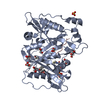
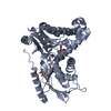
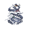
| Title | Crystal structure of the dengue virus serotype 2 envelope protein domain III in complex with the variable domains of Mab 4E11 | ||||||
 Components Components |
| ||||||
 Keywords Keywords | IMMUNE SYSTEM / dengue antibody neutralization | ||||||
| Function / homology |  Function and homology information Function and homology informationflavivirin / symbiont-mediated suppression of host JAK-STAT cascade via inhibition of host TYK2 activity / host cell mitochondrion / symbiont-mediated suppression of host JAK-STAT cascade via inhibition of STAT2 activity / symbiont-mediated suppression of host cytoplasmic pattern recognition receptor signaling pathway via inhibition of MAVS activity / viral capsid / nucleoside-triphosphate phosphatase / double-stranded RNA binding / channel activity / monoatomic ion transmembrane transport ...flavivirin / symbiont-mediated suppression of host JAK-STAT cascade via inhibition of host TYK2 activity / host cell mitochondrion / symbiont-mediated suppression of host JAK-STAT cascade via inhibition of STAT2 activity / symbiont-mediated suppression of host cytoplasmic pattern recognition receptor signaling pathway via inhibition of MAVS activity / viral capsid / nucleoside-triphosphate phosphatase / double-stranded RNA binding / channel activity / monoatomic ion transmembrane transport / clathrin-dependent endocytosis of virus by host cell / mRNA (guanine-N7)-methyltransferase / methyltransferase cap1 / molecular adaptor activity / methyltransferase cap1 activity / mRNA 5'-cap (guanine-N7-)-methyltransferase activity / RNA helicase activity / protein dimerization activity / host cell perinuclear region of cytoplasm / host cell endoplasmic reticulum membrane / RNA helicase / symbiont-mediated suppression of host type I interferon-mediated signaling pathway / serine-type endopeptidase activity / symbiont-mediated activation of host autophagy / RNA-directed RNA polymerase / viral RNA genome replication / RNA-directed RNA polymerase activity / fusion of virus membrane with host endosome membrane / viral envelope / lipid binding / virion attachment to host cell / host cell nucleus / virion membrane / structural molecule activity / ATP hydrolysis activity / proteolysis / extracellular region / ATP binding / metal ion binding Similarity search - Function | ||||||
| Biological species |  Dengue virus 2 Dengue virus 2 | ||||||
| Method |  X-RAY DIFFRACTION / SYNCHROTRON / MOLECULAR REPLACEMENT / Resolution: 2.1 Å X-RAY DIFFRACTION / SYNCHROTRON / MOLECULAR REPLACEMENT / Resolution: 2.1 Å | ||||||
 Authors Authors | Cockburn, J.J.B. / Navarro Sanchez, M.E. / Fretes, N. / Urvoas, A. / Staropoli, I. / Kikuti, C.M. / Coffey, L.L. / Arenzana Seisdedos, F. / Bedouelle, H. / Rey, F.A. | ||||||
 Citation Citation | Journal: Structure / Year: 2012 Title: Mechanism of dengue virus broad cross-neutralization by a monoclonal antibody. Authors: Cockburn, J.J. / Navarro Sanchez, M.E. / Fretes, N. / Urvoas, A. / Staropoli, I. / Kikuti, C.M. / Coffey, L.L. / Arenzana Seisdedos, F. / Bedouelle, H. / Rey, F.A. | ||||||
| History |
|
- Structure visualization
| Structure viewer | Molecule: MolmilJmol/JSmol |
|---|
- Downloads & links
-Download
| PDBx/mmCIF format | 3uzv.cif.gz | 145.5 KB | Display | PDBx/mmCIF format |
|---|---|---|---|---|
| PDB format | pdb3uzv.ent.gz | 114.4 KB | Display | PDB format |
| PDBx/mmJSON format | 3uzv.json.gz | Tree view | PDBx/mmJSON format | |
| Others |  Other downloads Other downloads |
-Validation report
| Arichive directory | https://data.pdbj.org/pub/pdb/validation_reports/uz/3uzvftp://data.pdbj.org/pub/pdb/validation_reports/uz/3uzv | HTTPS FTP |
|---|
-Related structure data
| Related structure data | 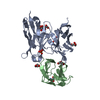 3uypC 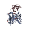 3uzeC 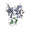 3uzqSC 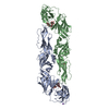 1oanS S: Starting model for refinement C: citing same article ( |
|---|---|
| Similar structure data |


-Links
 PDBj
PDBj


- Assembly
| Deposited unit | 
| ||||||||
|---|---|---|---|---|---|---|---|---|---|
| 1 |
| ||||||||
| Unit cell |
|
-Components
| #1: Protein | Mass: 12866.834 Da / Num. of mol.: 1 / Fragment: domain III (UNP residues 576-680) Source method: isolated from a genetically manipulated source Source: (gene. exp.) Dengue virus 2 / Strain: Jamaica/1409/1983 / Plasmid: pET20b / Production host:  |
|---|---|
| #2: Antibody | Mass: 27246.186 Da / Num. of mol.: 1 / Fragment: Single chain variable fragment Source method: isolated from a genetically manipulated source Source: (gene. exp.) |
| #3: Chemical | ChemComp-EOH /   Mass: 46.068 Da / Num. of mol.: 1 / Source method: obtained synthetically / Formula: C2H6O Mass: 46.068 Da / Num. of mol.: 1 / Source method: obtained synthetically / Formula: C2H6O |
| #4: Water | ChemComp-HOH /  Mass: 18.015 Da / Num. of mol.: 112 / Source method: isolated from a natural source / Formula: H2O Mass: 18.015 Da / Num. of mol.: 112 / Source method: isolated from a natural source / Formula: H2O |
| Has protein modification | Y |
-Experimental details
-Experiment
| Experiment | Method: X-RAY DIFFRACTION / Number of used crystals: 1 |
|---|
- Sample preparation
| Crystal | Density Matthews: 2.36 Å3/Da / Density % sol: 47.95 % |
|---|---|
| Crystal grow | Temperature: 292 K / Method: vapor diffusion, sitting drop / pH: 6.5 Details: JBS Classic 8 solution D1 (30% ethanol, 12% PEG 6K and 0.1M sodium acetate) diluted 70% in H20, pH 6.5, vapor diffusion, sitting drop, temperature 292K |
-Data collection
| Diffraction | Mean temperature: 100 K |
|---|---|
| Diffraction source | Source: SYNCHROTRON / Site: SLS  / Beamline: X06SA / Wavelength: 1.0721 Å / Beamline: X06SA / Wavelength: 1.0721 Å |
| Detector | Type: DECTRIS PILATUS 6M / Detector: PIXEL / Date: Jun 23, 2008 / Details: Dynamically bendable mirror |
| Radiation | Monochromator: LN2 cooled Fixed-exit Si(111) / Protocol: SINGLE WAVELENGTH / Monochromatic (M) / Laue (L): M / Scattering type: x-ray |
| Radiation wavelength | Wavelength: 1.0721 Å / Relative weight: 1 |
| Reflection | Resolution: 2.1→45.5 Å / Num. all: 22893 / Num. obs: 22893 / % possible obs: 98.3 % / Observed criterion σ(F): 0 / Observed criterion σ(I): -3 / Redundancy: 8.1 % / Biso Wilson estimate: 53.06 Å2 / Rmerge(I) obs: 0.046 / Net I/σ(I): 23.5 |
| Reflection shell | Resolution: 2.1→2.2 Å / Redundancy: 8.2 % / Rmerge(I) obs: 0.564 / Mean I/σ(I) obs: 3.3 / % possible all: 97.5 |
- Processing
| Software |
| ||||||||||||||||||||||||||||||||||||||||||||||||||||||||||||||||||||||||||||||||||||||||||||||||||||||||||||
|---|---|---|---|---|---|---|---|---|---|---|---|---|---|---|---|---|---|---|---|---|---|---|---|---|---|---|---|---|---|---|---|---|---|---|---|---|---|---|---|---|---|---|---|---|---|---|---|---|---|---|---|---|---|---|---|---|---|---|---|---|---|---|---|---|---|---|---|---|---|---|---|---|---|---|---|---|---|---|---|---|---|---|---|---|---|---|---|---|---|---|---|---|---|---|---|---|---|---|---|---|---|---|---|---|---|---|---|---|---|
| Refinement | Method to determine structure: MOLECULAR REPLACEMENT Starting model: 3UZQ, 1OAN Resolution: 2.1→45.52 Å / Cor.coef. Fo:Fc: 0.9427 / Cor.coef. Fo:Fc free: 0.9433 / Occupancy max: 1 / Occupancy min: 0 / SU R Cruickshank DPI: 0.202 / Cross valid method: THROUGHOUT / σ(F): 0 / Stereochemistry target values: Engh & Huber
| ||||||||||||||||||||||||||||||||||||||||||||||||||||||||||||||||||||||||||||||||||||||||||||||||||||||||||||
| Displacement parameters | Biso max: 173.58 Å2 / Biso mean: 60.9344 Å2 / Biso min: 36.15 Å2
| ||||||||||||||||||||||||||||||||||||||||||||||||||||||||||||||||||||||||||||||||||||||||||||||||||||||||||||
| Refine analyze | Luzzati coordinate error obs: 0.35 Å | ||||||||||||||||||||||||||||||||||||||||||||||||||||||||||||||||||||||||||||||||||||||||||||||||||||||||||||
| Refinement step | Cycle: LAST / Resolution: 2.1→45.52 Å
| ||||||||||||||||||||||||||||||||||||||||||||||||||||||||||||||||||||||||||||||||||||||||||||||||||||||||||||
| Refine LS restraints |
| ||||||||||||||||||||||||||||||||||||||||||||||||||||||||||||||||||||||||||||||||||||||||||||||||||||||||||||
| LS refinement shell | Resolution: 2.1→2.2 Å / Total num. of bins used: 11
| ||||||||||||||||||||||||||||||||||||||||||||||||||||||||||||||||||||||||||||||||||||||||||||||||||||||||||||
| Refinement TLS params. | Method: refined / Refine-ID: X-RAY DIFFRACTION
| ||||||||||||||||||||||||||||||||||||||||||||||||||||||||||||||||||||||||||||||||||||||||||||||||||||||||||||
| Refinement TLS group |
|
