 Movie
Movie Controller


+ Open data

- Basic information
| Entry | Database: PDB / ID: 1oan | |||||||||
|---|---|---|---|---|---|---|---|---|---|---|
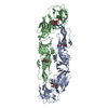
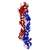
| Title | Crystal structure of the dengue 2 virus envelope protein | |||||||||
 Components Components | ENVELOPE GLYCOPROTEIN | |||||||||
 Keywords Keywords | GLYCOPROTEIN / DENGUE VIRUS / MEMBRANE FUSION / FLAVIVIRUS / FUSION PEPTIDE / LOW-PH CONFORMATIONAL CHANGE | |||||||||
| Function / homology |  Function and homology information Function and homology informationhost cell nucleolus / flavivirin / host cell mitochondrion / symbiont-mediated suppression of host JAK-STAT cascade via inhibition of host TYK2 activity / symbiont-mediated suppression of host JAK-STAT cascade via inhibition of STAT2 activity / symbiont-mediated suppression of host cytoplasmic pattern recognition receptor signaling pathway via inhibition of MAVS activity / viral capsid / nucleoside-triphosphate phosphatase / double-stranded RNA binding / channel activity ...host cell nucleolus / flavivirin / host cell mitochondrion / symbiont-mediated suppression of host JAK-STAT cascade via inhibition of host TYK2 activity / symbiont-mediated suppression of host JAK-STAT cascade via inhibition of STAT2 activity / symbiont-mediated suppression of host cytoplasmic pattern recognition receptor signaling pathway via inhibition of MAVS activity / viral capsid / nucleoside-triphosphate phosphatase / double-stranded RNA binding / channel activity / monoatomic ion transmembrane transport / clathrin-dependent endocytosis of virus by host cell / mRNA (guanine-N7)-methyltransferase / molecular adaptor activity / methyltransferase cap1 / methyltransferase cap1 activity / mRNA 5'-cap (guanine-N7-)-methyltransferase activity / RNA helicase activity / protein dimerization activity / host cell perinuclear region of cytoplasm / host cell endoplasmic reticulum membrane / RNA helicase / symbiont-mediated suppression of host type I interferon-mediated signaling pathway / serine-type endopeptidase activity / symbiont-mediated activation of host autophagy / RNA-directed RNA polymerase / viral RNA genome replication / RNA-directed RNA polymerase activity / fusion of virus membrane with host endosome membrane / viral envelope / lipid binding / virion attachment to host cell / virion membrane / structural molecule activity / DNA-templated transcription / ATP hydrolysis activity / proteolysis / extracellular region / ATP binding / metal ion binding Similarity search - Function | |||||||||
| Biological species |  DENGUE VIRUS TYPE 2 DENGUE VIRUS TYPE 2 | |||||||||
| Method |  X-RAY DIFFRACTION / SYNCHROTRON / MOLECULAR REPLACEMENT / Resolution: 2.75 Å X-RAY DIFFRACTION / SYNCHROTRON / MOLECULAR REPLACEMENT / Resolution: 2.75 Å | |||||||||
 Authors Authors | Modis, Y. / Harrison, S.C. | |||||||||
 Citation Citation | Journal: Proc.Natl.Acad.Sci.USA / Year: 2003 Title: A Ligand-Binding Pocket in the Dengue Virus Envelope Glycoprotein Authors: Modis, Y. / Ogata, S. / Clements, D. / Harrison, S.C. #1: Journal: Nature / Year: 1995Title: The Envelope Glycoprotein from Tick-Borne Encephalitis Virus at 2 A Resolution Authors: Rey, F.A. / Heinz, F.X. / Mandl, C. / Kunz, C. / Harrison, S.C. | |||||||||
| History |
| |||||||||
| Remark 650 | HELIX DETERMINATION METHOD: AUTHOR PROVIDED. | |||||||||
| Remark 700 | SHEET DETERMINATION METHOD: AUTHOR PROVIDED. |
- Structure visualization
| Structure viewer | Molecule: MolmilJmol/JSmol |
|---|
- Downloads & links
-Download
| PDBx/mmCIF format | 1oan.cif.gz | 169.8 KB | Display | PDBx/mmCIF format |
|---|---|---|---|---|
| PDB format | pdb1oan.ent.gz | 135.1 KB | Display | PDB format |
| PDBx/mmJSON format | 1oan.json.gz | Tree view | PDBx/mmJSON format | |
| Others |  Other downloads Other downloads |
-Validation report
| Arichive directory | https://data.pdbj.org/pub/pdb/validation_reports/oa/1oanftp://data.pdbj.org/pub/pdb/validation_reports/oa/1oan | HTTPS FTP |
|---|
-Related structure data
| Related structure data | 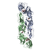 1okeC  1oam C: citing same article ( S: Starting model for refinement |
|---|---|
| Similar structure data |


-Links
 PDBj
PDBj


- Assembly
| Deposited unit | 
| ||||||||
|---|---|---|---|---|---|---|---|---|---|
| 1 |
| ||||||||
| Unit cell |
| ||||||||
| Noncrystallographic symmetry (NCS) | NCS oper: (Code: given Matrix: (0.979, -0.2, -0.038), Vector: |
-Components
| #1: Protein | Mass: 43819.391 Da / Num. of mol.: 2 / Fragment: SOLUBLE ECTODOMAIN, RESIDUES 281-674 Source method: isolated from a genetically manipulated source Source: (gene. exp.) DENGUE VIRUS TYPE 2 / Strain: STRAIN PR159/S1 / Plasmid: PMTT / Cell line (production host): SCHNEIDER 2 / Production host:  #2: Polysaccharide | Source method: isolated from a genetically manipulated source #3: Chemical | ChemComp-NA / |   Mass: 22.990 Da / Num. of mol.: 1 / Source method: obtained synthetically / Formula: Na Mass: 22.990 Da / Num. of mol.: 1 / Source method: obtained synthetically / Formula: Na#4: Sugar |   Type: D-saccharide, beta linking / Mass: 221.208 Da / Num. of mol.: 2 Type: D-saccharide, beta linking / Mass: 221.208 Da / Num. of mol.: 2Source method: isolated from a genetically manipulated source Formula: C8H15NO6 #5: Water | ChemComp-HOH / |  Mass: 18.015 Da / Num. of mol.: 45 / Source method: isolated from a natural source / Formula: H2O Mass: 18.015 Da / Num. of mol.: 45 / Source method: isolated from a natural source / Formula: H2OHas protein modification | Y | Sequence details | RESIDUES 100-108 FORM THE GLYCINE-RICH, HYDROPHOBIC FUSION PEPTIDE (ALLISON ET AL., J.VIROL. 75, ...RESIDUES 100-108 FORM THE GLYCINE-RICH, HYDROPHOBI | |
|---|
-Experimental details
-Experiment
| Experiment | Method: X-RAY DIFFRACTION / Number of used crystals: 1 |
|---|
- Sample preparation
| Crystal | Density Matthews: 3.395 Å3/Da / Density % sol: 63 % | ||||||||||||||||||||||||||||||||||||
|---|---|---|---|---|---|---|---|---|---|---|---|---|---|---|---|---|---|---|---|---|---|---|---|---|---|---|---|---|---|---|---|---|---|---|---|---|---|
| Crystal grow | pH: 8.5 Details: 11% PEG 8000, 1 M NACL, 0.1 M TRIS/HCL PH 9.0, 20% GLYCEROL | ||||||||||||||||||||||||||||||||||||
| Crystal grow | *PLUS Temperature: 4 ℃ / pH: 8 / Method: vapor diffusion, hanging drop | ||||||||||||||||||||||||||||||||||||
| Components of the solutions | *PLUS
|
-Data collection
| Diffraction | Mean temperature: 100 K |
|---|---|
| Diffraction source | Source: SYNCHROTRON / Site: CHESS  / Beamline: F1 / Wavelength: 0.91 / Beamline: F1 / Wavelength: 0.91 |
| Detector | Type: ADSC CCD / Detector: CCD / Date: Nov 15, 2002 |
| Radiation | Protocol: SINGLE WAVELENGTH / Monochromatic (M) / Laue (L): M / Scattering type: x-ray |
| Radiation wavelength | Wavelength: 0.91 Å / Relative weight: 1 |
| Reflection | Resolution: 2.75→50 Å / Num. obs: 27029 / % possible obs: 90.3 % / Redundancy: 10 % / Biso Wilson estimate: 34.3 Å2 / Rmerge(I) obs: 0.079 / Net I/σ(I): 21.7 |
| Reflection shell | Resolution: 2.75→2.85 Å / Redundancy: 1.4 % / Rmerge(I) obs: 0.409 / Mean I/σ(I) obs: 2.5 / % possible all: 48.8 |
| Reflection | *PLUS Highest resolution: 2.75 Å / Lowest resolution: 50 Å / % possible obs: 90 % / Rmerge(I) obs: 0.079 |
| Reflection shell | *PLUS % possible obs: 49 % / Rmerge(I) obs: 0.409 / Mean I/σ(I) obs: 2.5 |
- Processing
| Software |
| ||||||||||||||||||||||||||||||||||||||||||||||||||||||||||||||||||||||||||||||||
|---|---|---|---|---|---|---|---|---|---|---|---|---|---|---|---|---|---|---|---|---|---|---|---|---|---|---|---|---|---|---|---|---|---|---|---|---|---|---|---|---|---|---|---|---|---|---|---|---|---|---|---|---|---|---|---|---|---|---|---|---|---|---|---|---|---|---|---|---|---|---|---|---|---|---|---|---|---|---|---|---|---|
| Refinement | Method to determine structure: MOLECULAR REPLACEMENT Starting model: PDB ENTRY 1OAM 1oam Resolution: 2.75→48.1 Å / Rfactor Rfree error: 0.008 / Data cutoff high absF: 1691401.1 / Data cutoff low absF: 0 / Isotropic thermal model: RESTRAINED / Cross valid method: THROUGHOUT / σ(F): 0
| ||||||||||||||||||||||||||||||||||||||||||||||||||||||||||||||||||||||||||||||||
| Solvent computation | Solvent model: FLAT MODEL / Bsol: 49.8907 Å2 / ksol: 0.360391 e/Å3 | ||||||||||||||||||||||||||||||||||||||||||||||||||||||||||||||||||||||||||||||||
| Displacement parameters | Biso mean: 76.1 Å2
| ||||||||||||||||||||||||||||||||||||||||||||||||||||||||||||||||||||||||||||||||
| Refine analyze |
| ||||||||||||||||||||||||||||||||||||||||||||||||||||||||||||||||||||||||||||||||
| Refinement step | Cycle: LAST / Resolution: 2.75→48.1 Å
| ||||||||||||||||||||||||||||||||||||||||||||||||||||||||||||||||||||||||||||||||
| Refine LS restraints |
| ||||||||||||||||||||||||||||||||||||||||||||||||||||||||||||||||||||||||||||||||
| LS refinement shell | Resolution: 2.75→2.92 Å / Rfactor Rfree error: 0.049 / Total num. of bins used: 6
| ||||||||||||||||||||||||||||||||||||||||||||||||||||||||||||||||||||||||||||||||
| Xplor file |
| ||||||||||||||||||||||||||||||||||||||||||||||||||||||||||||||||||||||||||||||||
| Refinement | *PLUS Lowest resolution: 50 Å / σ(F): 0 / % reflection Rfree: 5 % / Rfactor obs: 0.261 / Rfactor Rfree: 0.296 / Rfactor Rwork: 0.261 | ||||||||||||||||||||||||||||||||||||||||||||||||||||||||||||||||||||||||||||||||
| Solvent computation | *PLUS | ||||||||||||||||||||||||||||||||||||||||||||||||||||||||||||||||||||||||||||||||
| Displacement parameters | *PLUS | ||||||||||||||||||||||||||||||||||||||||||||||||||||||||||||||||||||||||||||||||
| Refine LS restraints | *PLUS
| ||||||||||||||||||||||||||||||||||||||||||||||||||||||||||||||||||||||||||||||||
| LS refinement shell | *PLUS Rfactor Rfree: 0.54 / Rfactor Rwork: 0.478 |
