 Movie
Movie Controller


+ Open data

- Basic information
| Entry | Database: PDB / ID: 3s24 | ||||||
|---|---|---|---|---|---|---|---|
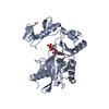
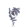
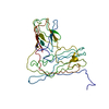
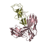
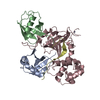
| Title | Crystal structure of human mRNA guanylyltransferase | ||||||
 Components Components | mRNA-capping enzyme | ||||||
 Keywords Keywords | HYDROLASE / TRANSFERASE / Capping enzyme / CE / HCE / GTASE / M7GPPPN CAP / GT/DNA ligase fold / transcription factor Spt5 / CTD | ||||||
| Function / homology |  Function and homology information Function and homology informationRNA guanylyltransferase activity / polynucleotide 5'-phosphatase activity / inorganic triphosphate phosphatase activity / mRNA 5'-phosphatase / mRNA 5'-triphosphate monophosphatase activity / RNA Pol II CTD phosphorylation and interaction with CE during HIV infection / RNA Pol II CTD phosphorylation and interaction with CE / mRNA Capping / 7-methylguanosine mRNA capping / RNA processing ...RNA guanylyltransferase activity / polynucleotide 5'-phosphatase activity / inorganic triphosphate phosphatase activity / mRNA 5'-phosphatase / mRNA 5'-triphosphate monophosphatase activity / RNA Pol II CTD phosphorylation and interaction with CE during HIV infection / RNA Pol II CTD phosphorylation and interaction with CE / mRNA Capping / 7-methylguanosine mRNA capping / RNA processing / phosphoprotein phosphatase activity / mRNA guanylyltransferase / mRNA guanylyltransferase activity / GTP binding / nucleoplasm / ATP binding / nucleus Similarity search - Function | ||||||
| Biological species |  Homo sapiens (human) Homo sapiens (human) | ||||||
| Method |  X-RAY DIFFRACTION / SYNCHROTRON / MOLECULAR REPLACEMENT / Resolution: 3.0137 Å X-RAY DIFFRACTION / SYNCHROTRON / MOLECULAR REPLACEMENT / Resolution: 3.0137 Å | ||||||
 Authors Authors | Das, K. / Chu, C. / Thyminski, J.R. / Bauman, J.D. / Guan, R. / Qiu, W. / Montelione, G.T. / Arnold, E. / Shatkin, A.J. | ||||||
 Citation Citation | Journal: Proc.Natl.Acad.Sci.USA / Year: 2011 Title: Structure of the guanylyltransferase domain of human mRNA capping enzyme. Authors: Chu, C. / Das, K. / Tyminski, J.R. / Bauman, J.D. / Guan, R. / Qiu, W. / Montelione, G.T. / Arnold, E. / Shatkin, A.J. | ||||||
| History |
|
- Structure visualization
| Structure viewer | Molecule: MolmilJmol/JSmol |
|---|
- Downloads & links
-Download
| PDBx/mmCIF format | 3s24.cif.gz | 450.8 KB | Display | PDBx/mmCIF format |
|---|---|---|---|---|
| PDB format | pdb3s24.ent.gz | 373.6 KB | Display | PDB format |
| PDBx/mmJSON format | 3s24.json.gz | Tree view | PDBx/mmJSON format | |
| Others |  Other downloads Other downloads |
-Validation report
| Arichive directory | https://data.pdbj.org/pub/pdb/validation_reports/s2/3s24ftp://data.pdbj.org/pub/pdb/validation_reports/s2/3s24 | HTTPS FTP |
|---|
-Related structure data
| Related structure data | 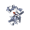 1cknS S: Starting model for refinement |
|---|---|
| Similar structure data |


-Links
 PDBj
PDBj


- Assembly
| Deposited unit | 
| ||||||||
|---|---|---|---|---|---|---|---|---|---|
| 1 | 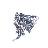
| ||||||||
| 2 | 
| ||||||||
| 3 | 
| ||||||||
| 4 | 
| ||||||||
| 5 | 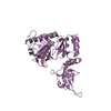
| ||||||||
| 6 | 
| ||||||||
| 7 | 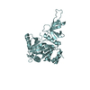
| ||||||||
| Unit cell |
|
-Components
| #1: Protein | Mass: 39988.984 Da / Num. of mol.: 7 / Fragment: UNP Residues 229-567 Source method: isolated from a genetically manipulated source Source: (gene. exp.) Homo sapiens (human) / Gene: RNGTT, CAP1A / Plasmid: pET20b / Production host:  References: UniProt: O60942, polynucleotide 5'-phosphatase, mRNA guanylyltransferase #2: Chemical | ChemComp-SO4 /   Mass: 96.063 Da / Num. of mol.: 5 / Source method: obtained synthetically / Formula: SO4 Mass: 96.063 Da / Num. of mol.: 5 / Source method: obtained synthetically / Formula: SO4 |
|---|
-Experimental details
-Experiment
| Experiment | Method: X-RAY DIFFRACTION |
|---|
- Sample preparation
| Crystal | Density Matthews: 2.57 Å3/Da / Density % sol: 52.07 % |
|---|---|
| Crystal grow | Temperature: 293 K / Method: vapor diffusion, hanging drop / pH: 6.5 Details: 1.7 M ammonium sulfate, 10% glucose, 5 mM 2-mercaptoethanol, 5 mM MgCl2, 5% isopropanol, pH 6.5, VAPOR DIFFUSION, HANGING DROP, temperature 293K |
-Data collection
| Diffraction | Mean temperature: 100 K |
|---|---|
| Diffraction source | Source: SYNCHROTRON / Site: NSLS  / Beamline: X25 / Wavelength: 0.9795 Å / Beamline: X25 / Wavelength: 0.9795 Å |
| Detector | Type: ADSC QUANTUM 315 / Detector: CCD / Date: Mar 10, 2009 |
| Radiation | Monochromator: tungsten-boron-carbide multilayer flms grown on silicon Protocol: SINGLE WAVELENGTH / Monochromatic (M) / Laue (L): M / Scattering type: x-ray |
| Radiation wavelength | Wavelength: 0.9795 Å / Relative weight: 1 |
| Reflection | Resolution: 3→50 Å / Num. obs: 53410 / % possible obs: 95.7 % / Observed criterion σ(F): 0 / Observed criterion σ(I): -1 / Redundancy: 3.5 % / Rmerge(I) obs: 0.085 / Net I/σ(I): 14.4 |
| Reflection shell | Resolution: 3→3.11 Å / Redundancy: 2.3 % / Rmerge(I) obs: 0.718 / Mean I/σ(I) obs: 1.2 / Num. unique all: 4128 / % possible all: 74.8 |
- Processing
| Software |
| |||||||||||||||||||||||||||||||||||||||||||||||||||||||||||||||||||||||||||||
|---|---|---|---|---|---|---|---|---|---|---|---|---|---|---|---|---|---|---|---|---|---|---|---|---|---|---|---|---|---|---|---|---|---|---|---|---|---|---|---|---|---|---|---|---|---|---|---|---|---|---|---|---|---|---|---|---|---|---|---|---|---|---|---|---|---|---|---|---|---|---|---|---|---|---|---|---|---|---|
| Refinement | Method to determine structure: MOLECULAR REPLACEMENT Starting model: PDB ENTRY 1CKN Resolution: 3.0137→35.427 Å / SU ML: 0.37 / σ(F): 0 / Phase error: 33.13 / Stereochemistry target values: Engh & Huber
| |||||||||||||||||||||||||||||||||||||||||||||||||||||||||||||||||||||||||||||
| Solvent computation | Shrinkage radii: 0.16 Å / VDW probe radii: 0.5 Å / Solvent model: FLAT BULK SOLVENT MODEL / Bsol: 33.474 Å2 / ksol: 0.35 e/Å3 | |||||||||||||||||||||||||||||||||||||||||||||||||||||||||||||||||||||||||||||
| Displacement parameters |
| |||||||||||||||||||||||||||||||||||||||||||||||||||||||||||||||||||||||||||||
| Refinement step | Cycle: LAST / Resolution: 3.0137→35.427 Å
| |||||||||||||||||||||||||||||||||||||||||||||||||||||||||||||||||||||||||||||
| Refine LS restraints |
| |||||||||||||||||||||||||||||||||||||||||||||||||||||||||||||||||||||||||||||
| LS refinement shell |
|
