 Movie
Movie Controller

[English] 日本語
 Yorodumi
- PDB-1ckm: STRUCTURE OF TWO DIFFERENT CONFORMATIONS OF MRNA CAPPING ENZYME I... -
+ Open data

- Basic information
| Entry | Database: PDB / ID: 1ckm | ||||||
|---|---|---|---|---|---|---|---|
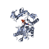
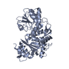
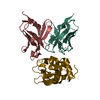
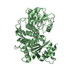
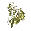
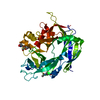
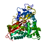
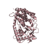
| Title | STRUCTURE OF TWO DIFFERENT CONFORMATIONS OF MRNA CAPPING ENZYME IN COMPLEX WITH GTP | ||||||
 Components Components | MRNA CAPPING ENZYME | ||||||
 Keywords Keywords | CAPPING ENZYME / MRNA / NUCLEOTIDYLTRANSFERASE | ||||||
| Function / homology |  Function and homology information Function and homology information7-methylguanosine mRNA capping / mRNA guanylyltransferase / mRNA guanylyltransferase activity / GTP binding / ATP binding Similarity search - Function | ||||||
| Biological species |  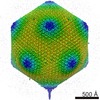 Paramecium bursaria Chlorella virus 1 Paramecium bursaria Chlorella virus 1 | ||||||
| Method |  X-RAY DIFFRACTION / SYNCHROTRON / MIR WITH A MERCURY DERIVATIVE OBTAINED BY SOAKING THE CRYSTAL FOR TWO HOURS IN 1 MM THIMEROSAL, A SELENOMETHIONINE DERIVATIVE / Resolution: 2.5 Å X-RAY DIFFRACTION / SYNCHROTRON / MIR WITH A MERCURY DERIVATIVE OBTAINED BY SOAKING THE CRYSTAL FOR TWO HOURS IN 1 MM THIMEROSAL, A SELENOMETHIONINE DERIVATIVE / Resolution: 2.5 Å | ||||||
 Authors Authors | Hakansson, K. / Doherty, A.J. / Wigley, D.B. | ||||||
 Citation Citation | Journal: Cell(Cambridge,Mass.) / Year: 1997 Title: X-ray crystallography reveals a large conformational change during guanyl transfer by mRNA capping enzymes. Authors: Hakansson, K. / Doherty, A.J. / Shuman, S. / Wigley, D.B. #1: Journal: To be PublishedTitle: Crystallization of the RNA Guanylyltransferase of Chlorella Virus Pbcv-1 Change During Guanyl Transfer by Mrna Capping Enzymes Authors: Doherty, A.J. / Hakansson, K. / Ho, C.K. / Shuman, S. / Wigley, D.B. | ||||||
| History |
|
- Structure visualization
| Structure viewer | Molecule: MolmilJmol/JSmol |
|---|
- Downloads & links
-Download
| PDBx/mmCIF format | 1ckm.cif.gz | 147.2 KB | Display | PDBx/mmCIF format |
|---|---|---|---|---|
| PDB format | pdb1ckm.ent.gz | 116.7 KB | Display | PDB format |
| PDBx/mmJSON format | 1ckm.json.gz | Tree view | PDBx/mmJSON format | |
| Others |  Other downloads Other downloads |
-Validation report
| Arichive directory | https://data.pdbj.org/pub/pdb/validation_reports/ck/1ckmftp://data.pdbj.org/pub/pdb/validation_reports/ck/1ckm | HTTPS FTP |
|---|
-Related structure data


-Links
 PDBj
PDBj
- Assembly
| Deposited unit | 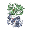
| ||||||||
|---|---|---|---|---|---|---|---|---|---|
| 1 | 
| ||||||||
| 2 | 
| ||||||||
| 3 | 
| ||||||||
| 4 |
| ||||||||
| 5 | 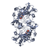
| ||||||||
| Unit cell |
| ||||||||
| Details | THE TWO MOLECULES IN THE ASYMMETRIC UNIT ARE IN DIFFERENT CONFORMATIONS. THE PROTEIN IS MONOMERIC AND THIS ENTRY MERELY REPRESENTS A CO-CRYSTAL OF THE ENZYME IN TWO DIFFERENT CONFORMATIONS. |
-Components
| #1: Protein | Mass: 37884.992 Da / Num. of mol.: 2 Source method: isolated from a genetically manipulated source Source: (gene. exp.) Paramecium bursaria Chlorella virus 1 / Genus: Chlorovirus / Production host:  #2: Chemical |   Mass: 523.180 Da / Num. of mol.: 2 / Source method: obtained synthetically / Formula: C10H16N5O14P3 / Comment: GTP, energy-carrying molecule*YM Mass: 523.180 Da / Num. of mol.: 2 / Source method: obtained synthetically / Formula: C10H16N5O14P3 / Comment: GTP, energy-carrying molecule*YM#3: Water | ChemComp-HOH / |  Mass: 18.015 Da / Num. of mol.: 360 / Source method: isolated from a natural source / Formula: H2O Mass: 18.015 Da / Num. of mol.: 360 / Source method: isolated from a natural source / Formula: H2O |
|---|
-Experimental details
-Experiment
| Experiment | Method: X-RAY DIFFRACTION / Number of used crystals: 1 |
|---|
- Sample preparation
| Crystal | Density Matthews: 3.5 Å3/Da / Density % sol: 65 % | ||||||||||||||||||||||||||||||||||||
|---|---|---|---|---|---|---|---|---|---|---|---|---|---|---|---|---|---|---|---|---|---|---|---|---|---|---|---|---|---|---|---|---|---|---|---|---|---|
| Crystal grow | Method: vapor diffusion, hanging drop / pH: 7.5 Details: HANGING DROP VAPOR DIFFUSION. 10-15 MG/ML PROTEIN IN 50 MM TRIS-HCL, 0.5 M NACL, 10 MM MGCL2, 5MM GTP, 2 MM EDTA, 4 MM DTT, PH 7.5 WERE MIXED WITH AN EQUAL VOLUME OF AND EQUILIBRATED AGAINST ...Details: HANGING DROP VAPOR DIFFUSION. 10-15 MG/ML PROTEIN IN 50 MM TRIS-HCL, 0.5 M NACL, 10 MM MGCL2, 5MM GTP, 2 MM EDTA, 4 MM DTT, PH 7.5 WERE MIXED WITH AN EQUAL VOLUME OF AND EQUILIBRATED AGAINST 100 MM TRIS-HCL, 200 MM NACL, 200 MM AMMONIUM SULFATE 34% MEOPEG 5000, PH 7.5., vapor diffusion - hanging drop | ||||||||||||||||||||||||||||||||||||
| Crystal grow | *PLUS Method: unknown / Details: Doherty, A.J., (1996) Nucl. Acids Res., 24. 2281. | ||||||||||||||||||||||||||||||||||||
| Components of the solutions | *PLUS
|
-Data collection
| Diffraction | Mean temperature: 100 K |
|---|---|
| Diffraction source | Source: SYNCHROTRON / Site: SRS  / Beamline: PX7.2 / Wavelength: 1.448 / Beamline: PX7.2 / Wavelength: 1.448 |
| Detector | Type: MAR scanner 300 mm plate / Detector: IMAGE PLATE / Date: Jul 1, 1996 |
| Radiation | Monochromatic (M) / Laue (L): M / Scattering type: x-ray |
| Radiation wavelength | Wavelength: 1.448 Å / Relative weight: 1 |
| Reflection | Resolution: 2.5→20 Å / Num. obs: 36196 / % possible obs: 97.5 % / Observed criterion σ(I): 0 / Redundancy: 3 % / Biso Wilson estimate: 48.3 Å2 / Rmerge(I) obs: 0.062 / Net I/σ(I): 13.3 |
- Processing
| Software |
| ||||||||||||||||||||||||||||||||||||||||||||||||||||||||||||
|---|---|---|---|---|---|---|---|---|---|---|---|---|---|---|---|---|---|---|---|---|---|---|---|---|---|---|---|---|---|---|---|---|---|---|---|---|---|---|---|---|---|---|---|---|---|---|---|---|---|---|---|---|---|---|---|---|---|---|---|---|---|
| Refinement | Method to determine structure: MIR WITH A MERCURY DERIVATIVE OBTAINED BY SOAKING THE CRYSTAL FOR TWO HOURS IN 1 MM THIMEROSAL, A SELENOMETHIONINE DERIVATIVE Resolution: 2.5→10 Å / σ(F): 0
| ||||||||||||||||||||||||||||||||||||||||||||||||||||||||||||
| Displacement parameters | Biso mean: 29.1 Å2 | ||||||||||||||||||||||||||||||||||||||||||||||||||||||||||||
| Refinement step | Cycle: LAST / Resolution: 2.5→10 Å
| ||||||||||||||||||||||||||||||||||||||||||||||||||||||||||||
| Refine LS restraints |
| ||||||||||||||||||||||||||||||||||||||||||||||||||||||||||||
| Xplor file |
| ||||||||||||||||||||||||||||||||||||||||||||||||||||||||||||
| Software | *PLUS Name: X-PLOR / Classification: refinement | ||||||||||||||||||||||||||||||||||||||||||||||||||||||||||||
| Refine LS restraints | *PLUS
|
