 Movie
Movie Controller

[English] 日本語
 Yorodumi
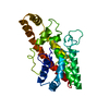
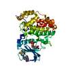
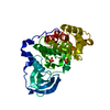
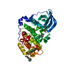
- PDB-1qm6: Closed form of Clostridium perfringens alpha-toxin strain NCTC8237 -
+ Open data

- Basic information
| Entry | Database: PDB / ID: 1qm6 | ||||||
|---|---|---|---|---|---|---|---|
| Title | Closed form of Clostridium perfringens alpha-toxin strain NCTC8237 | ||||||
 Components Components | PHOSPHOLIPASE C | ||||||
 Keywords Keywords | HYDROLASE / ZINC PHOSPHOLIPASE C / GANGRENE DETERMINANT / C2 DOMAIN / CA AND MEMBRANE BINDING. | ||||||
| Function / homology |  Function and homology information Function and homology information: / phospholipase C / phosphatidylcholine phospholipase C activity / hemolysis in another organism / symbiont-mediated killing of host cell / toxin activity / hydrolase activity / extracellular region / zinc ion binding Similarity search - Function | ||||||
| Biological species |   CLOSTRIDIUM PERFRINGENS (bacteria) CLOSTRIDIUM PERFRINGENS (bacteria) | ||||||
| Method |  X-RAY DIFFRACTION / SYNCHROTRON / MOLECULAR REPLACEMENT / Resolution: 2.5 Å X-RAY DIFFRACTION / SYNCHROTRON / MOLECULAR REPLACEMENT / Resolution: 2.5 Å | ||||||
 Authors Authors | Naylor, C.E. / Miller, J. / Titball, R.W. / Basak, A.K. | ||||||
 Citation Citation | Journal: J.Mol.Biol. / Year: 1999 Title: Characterisation of the Calcium-Binding C-Terminal Domain of Clostridium Perfringens Alpha-Toxin Authors: Naylor, C.E. / Jepson, M. / Crane, D.T. / Titball, R.W. / Miller, J. / Basak, A.K. / Bolgiano, B. #1: Journal: Nat.Struct.Biol. / Year: 1998Title: Structure of the Key Toxin in Gas-Gangrene Authors: Naylor, C.E. / Eaton, J.T. / Howells, A. / Justin, N. / Moss, D.S. / Titball, R.W. / Basak, A.K. #2: Journal: Acta Crystallogr.,Sect.D / Year: 1998 Title: Crystallisation and Preliminary X-Ray Diffraction Studies of Alpha-Toxin from Two Different Strains (Nctc-8237 and Cer89L43) of Clostridium Perfringens Authors: Basak, A.K. / Howells, A. / Eaton, J.T. / Moss, D.S. / Naylor, C. / Miller, J. / Titball, R.W. | ||||||
| History |
| ||||||
| Remark 650 | HELIX DETERMINATION METHOD: AUTHOR PROVIDED. | ||||||
| Remark 700 | SHEET DETERMINATION METHOD: AUTHOR PROVIDED. |
- Structure visualization
| Structure viewer | Molecule: MolmilJmol/JSmol |
|---|
- Downloads & links
-Download
| PDBx/mmCIF format | 1qm6.cif.gz | 159.8 KB | Display | PDBx/mmCIF format |
|---|---|---|---|---|
| PDB format | pdb1qm6.ent.gz | 126.1 KB | Display | PDB format |
| PDBx/mmJSON format | 1qm6.json.gz | Tree view | PDBx/mmJSON format | |
| Others |  Other downloads Other downloads |
-Validation report
| Arichive directory | https://data.pdbj.org/pub/pdb/validation_reports/qm/1qm6ftp://data.pdbj.org/pub/pdb/validation_reports/qm/1qm6 | HTTPS FTP |
|---|
-Related structure data
| Related structure data |  1qmdC  1ca1S S: Starting model for refinement C: citing same article ( |
|---|---|
| Similar structure data |


-Links
 PDBj
PDBj- Assembly
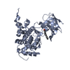
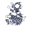
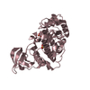
| Deposited unit | 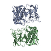
| ||||||||
|---|---|---|---|---|---|---|---|---|---|
| 1 | 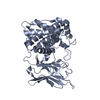
| ||||||||
| 2 | 
| ||||||||
| Unit cell |
| ||||||||
| Noncrystallographic symmetry (NCS) | NCS oper: (Code: given Matrix: (0.99865, -0.05189, -0.00206), Vector: |
-Components
| #1: Protein | Mass: 42577.816 Da / Num. of mol.: 2 Source method: isolated from a genetically manipulated source Source: (gene. exp.) CLOSTRIDIUM PERFRINGENS (bacteria) / Strain: NCTC8237 / Plasmid: PUC18 / Cellular location (production host): PERIPLASMIC / Gene (production host): CPA / Production host: References: UniProt: P15310, UniProt: Q0TV31*PLUS, phospholipase C #2: Chemical | ChemComp-ZN /   Mass: 65.409 Da / Num. of mol.: 4 / Source method: obtained synthetically / Formula: Zn Mass: 65.409 Da / Num. of mol.: 4 / Source method: obtained synthetically / Formula: Zn#3: Water | ChemComp-HOH / |  Mass: 18.015 Da / Num. of mol.: 89 / Source method: isolated from a natural source / Formula: H2O Mass: 18.015 Da / Num. of mol.: 89 / Source method: isolated from a natural source / Formula: H2OSequence details | 27 RESIDUES AT START OF SEQUENCE ARE A SIGNAL PEPTIDE NOT PRESENT IN MATURE, ACTIVE FOLDED PROTEIN. | |
|---|
-Experimental details
-Experiment
| Experiment | Method: X-RAY DIFFRACTION / Number of used crystals: 1 |
|---|
- Sample preparation
| Crystal | Density Matthews: 2.45 Å3/Da / Density % sol: 50 % | ||||||||||||||||||
|---|---|---|---|---|---|---|---|---|---|---|---|---|---|---|---|---|---|---|---|
| Crystal grow | Method: vapor diffusion, hanging drop / pH: 4.7 Details: PROTEIN WAS CRYSTALLISED BY HANGING DROP FROM 1.8-2.0 M NACL IN 0.1 M NA ACETATE, PH 4.7 OR 4.8 | ||||||||||||||||||
| Crystal grow | *PLUS Method: vapor diffusion, hanging drop | ||||||||||||||||||
| Components of the solutions | *PLUS
|
-Data collection
| Diffraction | Mean temperature: 100 K |
|---|---|
| Diffraction source | Source: SYNCHROTRON / Site: SRS  / Beamline: PX7.2 / Wavelength: 1.488 / Beamline: PX7.2 / Wavelength: 1.488 |
| Detector | Type: MARRESEARCH / Detector: IMAGE PLATE / Date: Jun 15, 1998 / Details: MIRRORS |
| Radiation | Monochromator: GE(111) / Protocol: SINGLE WAVELENGTH / Monochromatic (M) / Laue (L): M / Scattering type: x-ray |
| Radiation wavelength | Wavelength: 1.488 Å / Relative weight: 1 |
| Reflection | Resolution: 2.6→30 Å / Num. obs: 29145 / % possible obs: 90.5 % / Redundancy: 3.9 % / Biso Wilson estimate: 28.8 Å2 / Rmerge(I) obs: 0.121 / Net I/σ(I): 8.8 |
| Reflection shell | Resolution: 2.6→2.7 Å / Redundancy: 3.6 % / Rmerge(I) obs: 0.336 / Mean I/σ(I) obs: 4.2 / % possible all: 87.9 |
| Reflection | *PLUS Num. measured all: 98122 |
| Reflection shell | *PLUS % possible obs: 87.9 % / Num. unique obs: 3939 / Num. measured obs: 14192 |
- Processing
| Software |
| ||||||||||||||||||||||||||||||||||||||||||||||||||||||||||||||||||||||||||||||||
|---|---|---|---|---|---|---|---|---|---|---|---|---|---|---|---|---|---|---|---|---|---|---|---|---|---|---|---|---|---|---|---|---|---|---|---|---|---|---|---|---|---|---|---|---|---|---|---|---|---|---|---|---|---|---|---|---|---|---|---|---|---|---|---|---|---|---|---|---|---|---|---|---|---|---|---|---|---|---|---|---|---|
| Refinement | Method to determine structure: MOLECULAR REPLACEMENT Starting model: CALCIUM-FREE, CLOSED ALPHA-TOXIN STRAIN CER89L43 (PDB CODE: 1CA1) Resolution: 2.5→30 Å / Data cutoff high absF: 10000 / Isotropic thermal model: RESTRAINED / Cross valid method: THROUGHOUT / σ(F): 0
| ||||||||||||||||||||||||||||||||||||||||||||||||||||||||||||||||||||||||||||||||
| Solvent computation | Solvent model: FLAT MODEL (ANISOTROPIC) / Bsol: 10 Å2 / ksol: 0.406 e/Å3 | ||||||||||||||||||||||||||||||||||||||||||||||||||||||||||||||||||||||||||||||||
| Displacement parameters | Biso mean: 18.9 Å2
| ||||||||||||||||||||||||||||||||||||||||||||||||||||||||||||||||||||||||||||||||
| Refine analyze |
| ||||||||||||||||||||||||||||||||||||||||||||||||||||||||||||||||||||||||||||||||
| Refinement step | Cycle: LAST / Resolution: 2.5→30 Å
| ||||||||||||||||||||||||||||||||||||||||||||||||||||||||||||||||||||||||||||||||
| Refine LS restraints |
| ||||||||||||||||||||||||||||||||||||||||||||||||||||||||||||||||||||||||||||||||
| Refine LS restraints NCS | Rms dev Biso : 1.95 Å2 / Rms dev position: 0.031 Å / Weight Biso : 2 / Weight position: 300 | ||||||||||||||||||||||||||||||||||||||||||||||||||||||||||||||||||||||||||||||||
| LS refinement shell | Resolution: 2.5→2.6 Å / Total num. of bins used: 10
| ||||||||||||||||||||||||||||||||||||||||||||||||||||||||||||||||||||||||||||||||
| Xplor file |
| ||||||||||||||||||||||||||||||||||||||||||||||||||||||||||||||||||||||||||||||||
| Software | *PLUS Name: CNS / Version: 0.5 / Classification: refinement | ||||||||||||||||||||||||||||||||||||||||||||||||||||||||||||||||||||||||||||||||
| Refinement | *PLUS Rfactor obs: 0.22 / Rfactor Rwork: 0.22 | ||||||||||||||||||||||||||||||||||||||||||||||||||||||||||||||||||||||||||||||||
| Solvent computation | *PLUS | ||||||||||||||||||||||||||||||||||||||||||||||||||||||||||||||||||||||||||||||||
| Displacement parameters | *PLUS | ||||||||||||||||||||||||||||||||||||||||||||||||||||||||||||||||||||||||||||||||
| Refine LS restraints | *PLUS
|
