 Movie
Movie Controller


+ Open data

- Basic information
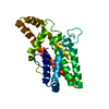
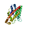
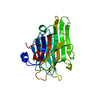
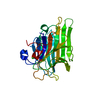
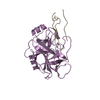
| Entry | Database: PDB / ID: 1ah7 | ||||||
|---|---|---|---|---|---|---|---|
| Title | PHOSPHOLIPASE C FROM BACILLUS CEREUS | ||||||
 Components Components | PHOSPHOLIPASE C | ||||||
 Keywords Keywords | HYDROLASE / LIPASE / PHOSPHOLIPID HYDROLYSIS | ||||||
| Function / homology |  Function and homology information Function and homology informationphospholipase C / phosphatidylcholine phospholipase C activity / killing of cells of another organism / zinc ion binding Similarity search - Function | ||||||
| Biological species |  | ||||||
| Method |  X-RAY DIFFRACTION / SYNCHROTRON / MIR / Resolution: 1.501 Å X-RAY DIFFRACTION / SYNCHROTRON / MIR / Resolution: 1.501 Å | ||||||
 Authors Authors | Greaves, R. | ||||||
 Citation Citation | Journal: Nature / Year: 1989 Title: High-resolution (1.5 A) crystal structure of phospholipase C from Bacillus cereus. Authors: Hough, E. / Hansen, L.K. / Birknes, B. / Jynge, K. / Hansen, S. / Hordvik, A. / Little, C. / Dodson, E. / Derewenda, Z. #1: Journal: Lipases : Their Structure, Biochemistry, and ApplicationYear: 1994 Title: Structural Aspects of Phospholipase C from Bacillus Cereus and its Reaction Mechanism Authors: Hough, E. / Hansen, S. #2: Journal: J.Mol.Biol. / Year: 1993Title: Crystal Structure of Phospholipase C from Bacillus Cereus Complexed with a Substrate Analog Authors: Hansen, S. / Hough, E. / Svensson, L.A. / Wong, Y.L. / Martin, S.F. #3: Journal: J.Mol.Biol. / Year: 1993Title: The Crystal Structure of Tris-Inhibited Phospholipase C from Bacillus Cereus at 1.9 A Resolution. The Nature of the Metal Ion in Site 2 Authors: Hansen, S. / Hansen, L.K. / Hough, E. #4: Journal: J.Mol.Biol. / Year: 1992Title: Crystal Structures of Phosphate, Iodide and Iodate-Inhibited Phospholipase C from Bacillus Cereus and Structural Investigations of the Binding of Reaction Products and a Substrate Analogue Authors: Hansen, S. / Hansen, L.K. / Hough, E. | ||||||
| History |
|
- Structure visualization
| Structure viewer | Molecule: MolmilJmol/JSmol |
|---|
- Downloads & links
-Download
| PDBx/mmCIF format | 1ah7.cif.gz | 68.4 KB | Display | PDBx/mmCIF format |
|---|---|---|---|---|
| PDB format | pdb1ah7.ent.gz | 51 KB | Display | PDB format |
| PDBx/mmJSON format | 1ah7.json.gz | Tree view | PDBx/mmJSON format | |
| Others |  Other downloads Other downloads |
-Validation report
| Arichive directory | https://data.pdbj.org/pub/pdb/validation_reports/ah/1ah7ftp://data.pdbj.org/pub/pdb/validation_reports/ah/1ah7 | HTTPS FTP |
|---|
-Related structure data
| Similar structure data |
|---|


-Links
 PDBj
PDBj- Assembly
| Deposited unit | 
| ||||||||
|---|---|---|---|---|---|---|---|---|---|
| 1 |
| ||||||||
| Unit cell |
|
-Components
| #1: Protein | Mass: 28423.420 Da / Num. of mol.: 1 / Source method: isolated from a natural source / Source: (natural) | ||
|---|---|---|---|
| #2: Chemical |   Mass: 65.409 Da / Num. of mol.: 3 / Source method: obtained synthetically / Formula: Zn Mass: 65.409 Da / Num. of mol.: 3 / Source method: obtained synthetically / Formula: Zn#3: Water | ChemComp-HOH / |  Mass: 18.015 Da / Num. of mol.: 232 / Source method: isolated from a natural source / Formula: H2O Mass: 18.015 Da / Num. of mol.: 232 / Source method: isolated from a natural source / Formula: H2O |
-Experimental details
-Experiment
| Experiment | Method: X-RAY DIFFRACTION / Number of used crystals: 2 |
|---|
- Sample preparation
| Crystal | Density Matthews: 2.62 Å3/Da / Density % sol: 52.7 % | ||||||||||||||||||||
|---|---|---|---|---|---|---|---|---|---|---|---|---|---|---|---|---|---|---|---|---|---|
| Crystal grow | Method: vapor diffusion / pH: 7 Details: VAPOR PHASE DIFFUSION FROM SOLUTION OF 5MG/ML PLC IN 35% SATURATED AMMONIUM SULFATE, pH 7, vapor diffusion | ||||||||||||||||||||
| Crystal grow | *PLUS Method: vapor diffusion | ||||||||||||||||||||
| Components of the solutions | *PLUS
|
-Data collection
| Diffraction | Mean temperature: 300 K |
|---|---|
| Diffraction source | Source: SYNCHROTRON / Site: SRS  / Beamline: PX9.6 / Wavelength: 0.87 / Beamline: PX9.6 / Wavelength: 0.87 |
| Detector | Type: ENRAF-NONIUS FAST / Detector: DIFFRACTOMETER / Date: Aug 1, 1986 / Details: MIRRORS |
| Radiation | Monochromator: GRAPHITE(002) / Monochromatic (M) / Laue (L): M / Scattering type: x-ray |
| Radiation wavelength | Wavelength: 0.87 Å / Relative weight: 1 |
| Reflection | Resolution: 1.5→12.45 Å / Num. obs: 46061 / % possible obs: 95 % / Redundancy: 2.5 % / Rsym value: 0.056 |
| Reflection shell | Resolution: 1.501→1.57 Å / % possible all: 82 |
| Reflection | *PLUS Rmerge(I) obs: 0.056 |
| Reflection shell | *PLUS % possible obs: 82 % |
- Processing
| Software |
| ||||||||||||||||
|---|---|---|---|---|---|---|---|---|---|---|---|---|---|---|---|---|---|
| Refinement | Method to determine structure: MIR / Resolution: 1.501→12.451 Å / Cross valid method: FREE R / σ(F): 0 Details: PROTEIN MODEL DID NOT INCLUDE HYDROGEN ATOM POSITIONS. INITIAL REFINEMENT WAS PERFORMED USING USING PROTIN AND PROLSQ. THERE IS STILL SOME DOUBT ABOUT THE EXACT POSITIONING OF TYR 156 WHICH ...Details: PROTEIN MODEL DID NOT INCLUDE HYDROGEN ATOM POSITIONS. INITIAL REFINEMENT WAS PERFORMED USING USING PROTIN AND PROLSQ. THERE IS STILL SOME DOUBT ABOUT THE EXACT POSITIONING OF TYR 156 WHICH IS NOT WITHIN THE EXPECTED DISTRIBUTION OF RING PLANARITY FOR TYROSINE RESIDUES. THE DENSITY AROUND THE C-TERMINUS WAS POOR. THE POSITIONING OF RESIDUES ASP 244 AND ARG 245 IS THUS AMBIGUOUS.
| ||||||||||||||||
| Refinement step | Cycle: LAST / Resolution: 1.501→12.451 Å
| ||||||||||||||||
| Software | *PLUS Name: REFMAC / Classification: refinement | ||||||||||||||||
| Refinement | *PLUS Num. reflection all: 48722 | ||||||||||||||||
| Solvent computation | *PLUS | ||||||||||||||||
| Displacement parameters | *PLUS | ||||||||||||||||
| Refine LS restraints | *PLUS
|
