negative regulation of glycogen (starch) synthase activity / neuron projection organization / regulation of microtubule anchoring at centrosome / negative regulation of mesenchymal stem cell differentiation / negative regulation of type B pancreatic cell development / superior temporal gyrus development / positive regulation of protein localization to cilium / negative regulation of glycogen biosynthetic process / negative regulation of TORC2 signaling / beta-arrestin-dependent dopamine receptor signaling pathway ...negative regulation of glycogen (starch) synthase activity / neuron projection organization / regulation of microtubule anchoring at centrosome / negative regulation of mesenchymal stem cell differentiation / negative regulation of type B pancreatic cell development / superior temporal gyrus development / positive regulation of protein localization to cilium / negative regulation of glycogen biosynthetic process / negative regulation of TORC2 signaling / beta-arrestin-dependent dopamine receptor signaling pathway / negative regulation of dopaminergic neuron differentiation / positive regulation of protein localization to centrosome / maintenance of cell polarity / positive regulation of cilium assembly / CRMPs in Sema3A signaling / tau-protein kinase / heart valve development / beta-catenin destruction complex / APC truncation mutants have impaired AXIN binding / AXIN missense mutants destabilize the destruction complex / Truncations of AMER1 destabilize the destruction complex / positive regulation of mitochondrial outer membrane permeabilization involved in apoptotic signaling pathway / regulation of protein export from nucleus / cellular response to interleukin-3 / Maturation of nucleoprotein / Beta-catenin phosphorylation cascade / Signaling by GSK3beta mutants / CTNNB1 S33 mutants aren't phosphorylated / CTNNB1 S37 mutants aren't phosphorylated / CTNNB1 S45 mutants aren't phosphorylated / CTNNB1 T41 mutants aren't phosphorylated / regulation of long-term synaptic potentiation / Wnt signalosome / negative regulation of TOR signaling / regulation of microtubule-based process / AKT phosphorylates targets in the cytosol / Disassembly of the destruction complex and recruitment of AXIN to the membrane / positive regulation of protein binding / regulation of axon extension / negative regulation of calcineurin-NFAT signaling cascade / Maturation of nucleoprotein / negative regulation of protein localization to nucleus / negative regulation of epithelial to mesenchymal transition / tau-protein kinase activity / positive regulation of cell-matrix adhesion / glycogen metabolic process / ER overload response / regulation of axonogenesis / regulation of dendrite morphogenesis / regulation of neuron projection development / Constitutive Signaling by AKT1 E17K in Cancer / protein kinase A catalytic subunit binding / establishment of cell polarity / dynactin binding / epithelial to mesenchymal transition / Regulation of HSF1-mediated heat shock response / canonical Wnt signaling pathway / negative regulation of osteoblast differentiation / NF-kappaB binding / negative regulation of extrinsic apoptotic signaling pathway via death domain receptors / negative regulation of protein-containing complex assembly / regulation of cellular response to heat / extrinsic apoptotic signaling pathway / cellular response to retinoic acid / extrinsic apoptotic signaling pathway in absence of ligand / positive regulation of type I interferon production / Transcriptional and post-translational regulation of MITF-M expression and activity / positive regulation of autophagy / response to endoplasmic reticulum stress / positive regulation of protein export from nucleus / presynaptic modulation of chemical synaptic transmission / negative regulation of cell migration / peptidyl-serine phosphorylation / positive regulation of protein ubiquitination / regulation of microtubule cytoskeleton organization / hippocampus development / positive regulation of cell differentiation / mitochondrion organization / Ubiquitin-dependent degradation of Cyclin D / excitatory postsynaptic potential / positive regulation of protein-containing complex assembly / negative regulation of canonical Wnt signaling pathway / circadian rhythm / regulation of circadian rhythm / GSK3B and BTRC:CUL1-mediated-degradation of NFE2L2 / beta-catenin binding / Degradation of GLI2 by the proteasome / GLI3 is processed to GLI3R by the proteasome / B-WICH complex positively regulates rRNA expression / Degradation of beta-catenin by the destruction complex / tau protein binding / cellular response to amyloid-beta / Wnt signaling pathway / neuron projection development / kinase activity / p53 binding / positive regulation of protein catabolic process / Regulation of RUNX2 expression and activity / insulin receptor signaling pathway / protein autophosphorylation Similarity search - Function
Glycogen synthase kinase 3, catalytic domain / : / Phosphorylase Kinase; domain 1 / Phosphorylase Kinase; domain 1 / Transferase(Phosphotransferase) domain 1 / Transferase(Phosphotransferase); domain 1 / Serine/threonine-protein kinase, active site / Serine/Threonine protein kinases active-site signature. / Protein kinase domain / Serine/Threonine protein kinases, catalytic domain ...Glycogen synthase kinase 3, catalytic domain / : / Phosphorylase Kinase; domain 1 / Phosphorylase Kinase; domain 1 / Transferase(Phosphotransferase) domain 1 / Transferase(Phosphotransferase); domain 1 / Serine/threonine-protein kinase, active site / Serine/Threonine protein kinases active-site signature. / Protein kinase domain / Serine/Threonine protein kinases, catalytic domain / Protein kinase, ATP binding site / Protein kinases ATP-binding region signature. / Protein kinase domain profile. / Protein kinase domain / Protein kinase-like domain superfamily / 2-Layer Sandwich / Orthogonal Bundle / Mainly Alpha / Alpha Beta Similarity search - Domain/homology
SHEET DETERMINATION METHOD: DSSP THE SHEETS PRESENTED AS "AA" IN EACH CHAIN ON SHEET RECORDS BELOW ... SHEET DETERMINATION METHOD: DSSP THE SHEETS PRESENTED AS "AA" IN EACH CHAIN ON SHEET RECORDS BELOW IS ACTUALLY AN 6-STRANDED BARREL THIS IS REPRESENTED BY A 7-STRANDED SHEET IN WHICH THE FIRST AND LAST STRANDS ARE IDENTICAL. THE SHEETS PRESENTED AS "BA" IN EACH CHAIN ON SHEET RECORDS BELOW IS ACTUALLY AN 6-STRANDED BARREL THIS IS REPRESENTED BY A 7-STRANDED SHEET IN WHICH THE FIRST AND LAST STRANDS ARE IDENTICAL.
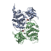
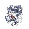
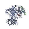
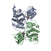
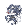
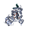
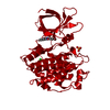
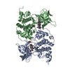
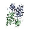
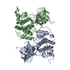
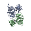
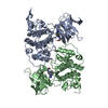
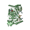
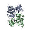
Mass: 52074.867 Da / Num. of mol.: 2 Source method: isolated from a genetically manipulated source Source: (gene. exp.) HOMO SAPIENS (human) / Production host: TRICHOPLUSIA NI (cabbage looper) References: UniProt: P49841, non-specific serine/threonine protein kinase, tau-protein kinase
Mass: 18.015 Da / Num. of mol.: 166 / Source method: isolated from a natural source / Formula: H2O
-
Experimental details
-
Experiment
Experiment
Method: X-RAY DIFFRACTION
-
Sample preparation
Crystal
Density Matthews: 3.06 Å3/Da / Density % sol: 59.8 % Description: THE EFFECTIVE RESOLUTION IS LOWER THAN 2.6 A, BUT THE WEAK DATA UPTO 2.6 A WERE INCLUDED IN THE REFINEMENT
Protocol: SINGLE WAVELENGTH / Monochromatic (M) / Laue (L): M / Scattering type: x-ray
Radiation wavelength
Wavelength: 1.5418 Å / Relative weight: 1
Reflection
Resolution: 2.6→20 Å / Num. obs: 38939 / % possible obs: 97.6 % / Observed criterion σ(I): 1.1 / Redundancy: 2.7 % / Biso Wilson estimate: 65.09 Å2 / Rmerge(I) obs: 0.15 / Net I/σ(I): 5.4
Reflection shell
Resolution: 2.6→2.74 Å / Redundancy: 2.7 % / Rmerge(I) obs: 0.78 / Mean I/σ(I) obs: 1.1 / % possible all: 99.5
-
Processing
Software
Name
Version
Classification
BUSTER
2.11.1
refinement
MOSFLM
datareduction
SCALA
datascaling
Refinement
Method to determine structure: MOLECULAR REPLACEMENT / Resolution: 2.6→19.94 Å / Cor.coef. Fo:Fc: 0.8797 / Cor.coef. Fo:Fc free: 0.8565 / SU R Cruickshank DPI: 0.371 / Cross valid method: THROUGHOUT / σ(F): 0 / SU R Blow DPI: 0.37 / SU Rfree Blow DPI: 0.253 / SU Rfree Cruickshank DPI: 0.256 Details: IDEAL-DIST CONTACT TERM CONTACT SETUP. ALL ATOMS HAVE CCP4 ATOM TYPE FROM LIBRARY
Rfactor
Num. reflection
% reflection
Selection details
Rfree
0.2517
1556
4.01 %
RANDOM
Rwork
0.2304
-
-
-
obs
0.2313
38823
97.05 %
-
Displacement parameters
Biso mean: 86.58 Å2
Baniso -1
Baniso -2
Baniso -3
1-
13.0999 Å2
0 Å2
0 Å2
2-
-
-39.3304 Å2
0 Å2
3-
-
-
26.2305 Å2
Refine analyze
Luzzati coordinate error obs: 0.486 Å
Refinement step
Cycle: LAST / Resolution: 2.6→19.94 Å
Protein
Nucleic acid
Ligand
Solvent
Total
Num. atoms
5610
0
62
166
5838
Refine LS restraints
Refine-ID
Type
Dev ideal
Number
Restraint function
Weight
X-RAY DIFFRACTION
t_bond_d
0.01
5818
HARMONIC
2
X-RAY DIFFRACTION
t_angle_deg
1.15
7918
HARMONIC
2
X-RAY DIFFRACTION
t_dihedral_angle_d
1988
SINUSOIDAL
2
X-RAY DIFFRACTION
t_incorr_chiral_ct
X-RAY DIFFRACTION
t_pseud_angle
X-RAY DIFFRACTION
t_trig_c_planes
126
HARMONIC
2
X-RAY DIFFRACTION
t_gen_planes
838
HARMONIC
5
X-RAY DIFFRACTION
t_it
5818
HARMONIC
20
X-RAY DIFFRACTION
t_nbd
X-RAY DIFFRACTION
t_omega_torsion
2.78
X-RAY DIFFRACTION
t_other_torsion
20.09
X-RAY DIFFRACTION
t_improper_torsion
X-RAY DIFFRACTION
t_chiral_improper_torsion
750
SEMIHARMONIC
5
X-RAY DIFFRACTION
t_sum_occupancies
X-RAY DIFFRACTION
t_utility_distance
X-RAY DIFFRACTION
t_utility_angle
X-RAY DIFFRACTION
t_utility_torsion
X-RAY DIFFRACTION
t_ideal_dist_contact
6470
SEMIHARMONIC
4
LS refinement shell
Resolution: 2.6→2.67 Å / Total num. of bins used: 19
Rfactor
Num. reflection
% reflection
Rfree
0.2951
120
3.96 %
Rwork
0.262
2910
-
all
0.2634
3030
-
obs
-
-
97.05 %
+
About Yorodumi
-
News
-
Feb 9, 2022. New format data for meta-information of EMDB entries
New format data for meta-information of EMDB entries
Version 3 of the EMDB header file is now the official format.
The previous official version 1.9 will be removed from the archive.
In the structure databanks used in Yorodumi, some data are registered as the other names, "COVID-19 virus" and "2019-nCoV". Here are the details of the virus and the list of structure data.
Jan 31, 2019. EMDB accession codes are about to change! (news from PDBe EMDB page)
EMDB accession codes are about to change! (news from PDBe EMDB page)
The allocation of 4 digits for EMDB accession codes will soon come to an end. Whilst these codes will remain in use, new EMDB accession codes will include an additional digit and will expand incrementally as the available range of codes is exhausted. The current 4-digit format prefixed with “EMD-” (i.e. EMD-XXXX) will advance to a 5-digit format (i.e. EMD-XXXXX), and so on. It is currently estimated that the 4-digit codes will be depleted around Spring 2019, at which point the 5-digit format will come into force.
The EM Navigator/Yorodumi systems omit the EMD- prefix.
Related info.:Q: What is EMD? / ID/Accession-code notation in Yorodumi/EM Navigator
Yorodumi is a browser for structure data from EMDB, PDB, SASBDB, etc.
This page is also the successor to EM Navigator detail page, and also detail information page/front-end page for Omokage search.
The word "yorodu" (or yorozu) is an old Japanese word meaning "ten thousand". "mi" (miru) is to see.
Related info.:EMDB / PDB / SASBDB / Comparison of 3 databanks / Yorodumi Search / Aug 31, 2016. New EM Navigator & Yorodumi / Yorodumi Papers / Jmol/JSmol / Function and homology information / Changes in new EM Navigator and Yorodumi
 Movie
Movie Controller


 Open data
 Basic information
 Components
 Keywords
 Function and homology information
 HOMO SAPIENS (human)
 X-RAY DIFFRACTION /
  Authors
 Citation
 Structure visualization
 Downloads & links
 Other downloads


 PDBj
PDBj


 Assembly
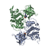
 TRICHOPLUSIA NI (cabbage looper)
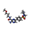
 Mass: 448.539 Da / Num. of mol.: 2 / Source method: obtained synthetically / Formula: C20H28N6O4S
 Mass: 18.015 Da / Num. of mol.: 166 / Source method: isolated from a natural source / Formula: H2O
 Sample preparation
 Processing