 Movie
Movie Controller

[English] 日本語
 Yorodumi
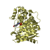
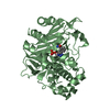
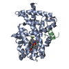
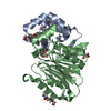
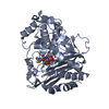
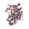
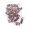
- PDB-1o6k: Structure of activated form of PKB kinase domain S474D with GSK3 ... -
+ Open data

- Basic information
| Entry | Database: PDB / ID: 1o6k | ||||||
|---|---|---|---|---|---|---|---|
| Title | Structure of activated form of PKB kinase domain S474D with GSK3 peptide and AMP-PNP | ||||||
 Components Components |
| ||||||
 Keywords Keywords | TRANSFERASE / PROTEIN KINASE / SERINE/THREONINE-PROTEIN KINASE | ||||||
| Function / homology |  Function and homology information Function and homology informationretinal rod cell apoptotic process / PDE3B signalling / cellular response to high light intensity / Inhibition of TSC complex formation by AKT (PKB) / positive regulation of cap-dependent translational initiation / AKT-mediated inactivation of FOXO1A / negative regulation of long-chain fatty acid import across plasma membrane / Negative regulation of the PI3K/AKT network / Activation of AKT2 / AKT phosphorylates targets in the nucleus ...retinal rod cell apoptotic process / PDE3B signalling / cellular response to high light intensity / Inhibition of TSC complex formation by AKT (PKB) / positive regulation of cap-dependent translational initiation / AKT-mediated inactivation of FOXO1A / negative regulation of long-chain fatty acid import across plasma membrane / Negative regulation of the PI3K/AKT network / Activation of AKT2 / AKT phosphorylates targets in the nucleus / negative regulation of glycogen (starch) synthase activity / neuron projection organization / regulation of microtubule anchoring at centrosome / negative regulation of mesenchymal stem cell differentiation / negative regulation of type B pancreatic cell development / superior temporal gyrus development / positive regulation of protein localization to cilium / negative regulation of glycogen biosynthetic process / positive regulation of fatty acid beta-oxidation / positive regulation of glucose metabolic process / negative regulation of TORC2 signaling / RUNX2 regulates genes involved in cell migration / beta-arrestin-dependent dopamine receptor signaling pathway / negative regulation of dopaminergic neuron differentiation / positive regulation of protein localization to centrosome / maintenance of cell polarity / mammary gland epithelial cell differentiation / positive regulation of cilium assembly / CRMPs in Sema3A signaling / tau-protein kinase / heart valve development / beta-catenin destruction complex / RAB GEFs exchange GTP for GDP on RABs / APC truncation mutants have impaired AXIN binding / AXIN missense mutants destabilize the destruction complex / Truncations of AMER1 destabilize the destruction complex / glycogen biosynthetic process / peripheral nervous system myelin maintenance / positive regulation of mitochondrial outer membrane permeabilization involved in apoptotic signaling pathway / regulation of protein export from nucleus / cellular response to interleukin-3 / Maturation of nucleoprotein / Beta-catenin phosphorylation cascade / Signaling by GSK3beta mutants / CTNNB1 S33 mutants aren't phosphorylated / CTNNB1 S37 mutants aren't phosphorylated / CTNNB1 S45 mutants aren't phosphorylated / CTNNB1 T41 mutants aren't phosphorylated / regulation of long-term synaptic potentiation / Wnt signalosome / negative regulation of TOR signaling / regulation of microtubule-based process / AKT phosphorylates targets in the cytosol / Disassembly of the destruction complex and recruitment of AXIN to the membrane / positive regulation of protein binding / regulation of axon extension / negative regulation of calcineurin-NFAT signaling cascade / positive regulation of cell motility / Maturation of nucleoprotein / Regulation of TP53 Activity through Association with Co-factors / negative regulation of protein localization to nucleus / negative regulation of epithelial to mesenchymal transition / tau-protein kinase activity / positive regulation of cell-matrix adhesion / Co-inhibition by CTLA4 / glycogen metabolic process / ER overload response / regulation of axonogenesis / regulation of dendrite morphogenesis / regulation of neuron projection development / negative regulation of PERK-mediated unfolded protein response / Constitutive Signaling by AKT1 E17K in Cancer / protein kinase A catalytic subunit binding / establishment of cell polarity / Regulation of MITF-M-dependent genes involved in pigmentation / Regulation of localization of FOXO transcription factors / dynactin binding / epithelial to mesenchymal transition / Activation of BAD and translocation to mitochondria / CD28 dependent PI3K/Akt signaling / Regulation of HSF1-mediated heat shock response / Estrogen-dependent nuclear events downstream of ESR-membrane signaling / positive regulation of blood vessel endothelial cell migration / positive regulation of glycogen biosynthetic process / positive regulation of protein targeting to membrane / canonical Wnt signaling pathway / negative regulation of osteoblast differentiation / SARS-CoV-2 targets host intracellular signalling and regulatory pathways / NF-kappaB binding / fat cell differentiation / negative regulation of extrinsic apoptotic signaling pathway via death domain receptors / Cyclin E associated events during G1/S transition / Cyclin A:Cdk2-associated events at S phase entry / negative regulation of protein-containing complex assembly / regulation of cellular response to heat / Regulation of TP53 Activity through Acetylation / extrinsic apoptotic signaling pathway / cellular response to retinoic acid / extrinsic apoptotic signaling pathway in absence of ligand / positive regulation of type I interferon production Similarity search - Function | ||||||
| Biological species |  HOMO SAPIENS (human) HOMO SAPIENS (human) | ||||||
| Method |  X-RAY DIFFRACTION / SYNCHROTRON / MOLECULAR REPLACEMENT / Resolution: 1.7 Å X-RAY DIFFRACTION / SYNCHROTRON / MOLECULAR REPLACEMENT / Resolution: 1.7 Å | ||||||
 Authors Authors | Yang, J. / Cron, P. / Good, V.M. / Thompson, V. / Hemmings, B.A. / Barford, D. | ||||||
 Citation Citation | Journal: Nat.Struct.Biol. / Year: 2002 Title: Crystal Structure of an Activated Akt/Protein Kinase B Ternary Complex with Gsk-3 Peptide and AMP-Pnp Authors: Yang, J. / Cron, P. / Good, V.M. / Thompson, V. / Hemmings, B.A. / Barford, D. | ||||||
| History |
|
- Structure visualization
| Structure viewer | Molecule: MolmilJmol/JSmol |
|---|
- Downloads & links
-Download
| PDBx/mmCIF format | 1o6k.cif.gz | 89 KB | Display | PDBx/mmCIF format |
|---|---|---|---|---|
| PDB format | pdb1o6k.ent.gz | 65.7 KB | Display | PDB format |
| PDBx/mmJSON format | 1o6k.json.gz | Tree view | PDBx/mmJSON format | |
| Others |  Other downloads Other downloads |
-Validation report
| Arichive directory | https://data.pdbj.org/pub/pdb/validation_reports/o6/1o6kftp://data.pdbj.org/pub/pdb/validation_reports/o6/1o6k | HTTPS FTP |
|---|
-Related structure data


-Links
 PDBj
PDBj


- Assembly
| Deposited unit | 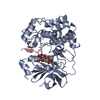
| ||||||||
|---|---|---|---|---|---|---|---|---|---|
| 1 |
| ||||||||
| Unit cell |
| ||||||||
| Details | CHAIN A IS A MONOMER, BUT THIS ENTRY IS CLASSIFIEDAS A DIMER AS CHAIN A IS IN COMPLEX WITH A PEPTIDE(CHAIN C), FORMING A DIMERIC ASSOCIATION. |
-Components
| #1: Protein | Mass: 39121.645 Da / Num. of mol.: 1 / Fragment: KINASE DOMAIN, RESIDUES 146-481 / Mutation: YES Source method: isolated from a genetically manipulated source Source: (gene. exp.) HOMO SAPIENS (human) / Production host:   SPODOPTERA FRUGIPERDA (fall armyworm) SPODOPTERA FRUGIPERDA (fall armyworm)References: UniProt: P31751, Transferases; Transferring phosphorus-containing groups; Phosphotransferases with an alcohol group as acceptor | ||||||
|---|---|---|---|---|---|---|---|
| #2: Protein/peptide | Mass: 1123.220 Da / Num. of mol.: 1 / Fragment: PEPTIDE, RESIDUES 3-12 Source method: isolated from a genetically manipulated source Source: (gene. exp.) HOMO SAPIENS (human) / Production host: SPODOPTERA FRUGIPERDA (fall armyworm) / References: UniProt: P49841, EC: 2.7.1.37 | ||||||
| #3: Chemical | ChemComp-ANP /   Mass: 506.196 Da / Num. of mol.: 1 / Source method: obtained synthetically / Formula: C10H17N6O12P3 / Comment: AMP-PNP, energy-carrying molecule analogue*YM Mass: 506.196 Da / Num. of mol.: 1 / Source method: obtained synthetically / Formula: C10H17N6O12P3 / Comment: AMP-PNP, energy-carrying molecule analogue*YM | ||||||
| #4: Chemical |   Mass: 54.938 Da / Num. of mol.: 2 / Source method: obtained synthetically / Formula: Mn Mass: 54.938 Da / Num. of mol.: 2 / Source method: obtained synthetically / Formula: Mn#5: Water | ChemComp-HOH / |  Mass: 18.015 Da / Num. of mol.: 281 / Source method: isolated from a natural source / Formula: H2O Mass: 18.015 Da / Num. of mol.: 281 / Source method: isolated from a natural source / Formula: H2OCompound details | PROTEIN KINASE KNOWN TO PHOSPHORYL | Has protein modification | Y | |
-Experimental details
-Experiment
| Experiment | Method: X-RAY DIFFRACTION / Number of used crystals: 1 |
|---|
- Sample preparation
| Crystal | Density Matthews: 2.6 Å3/Da / Density % sol: 49 % | ||||||||||||||||||||||||||||||||||||||||||||||||
|---|---|---|---|---|---|---|---|---|---|---|---|---|---|---|---|---|---|---|---|---|---|---|---|---|---|---|---|---|---|---|---|---|---|---|---|---|---|---|---|---|---|---|---|---|---|---|---|---|---|
| Crystal grow | pH: 7.5 Details: 10 MG/ML PROTEIN 20 PEG 4K, 10% ISOPROPONAL, 5 MM DTT, pH 7.50 | ||||||||||||||||||||||||||||||||||||||||||||||||
| Crystal grow | *PLUS Temperature: 20 ℃ / Method: unknown | ||||||||||||||||||||||||||||||||||||||||||||||||
| Components of the solutions | *PLUS
|
-Data collection
| Diffraction | Mean temperature: 100 K |
|---|---|
| Diffraction source | Source: SYNCHROTRON / Site: ESRF  / Beamline: ID14-4 / Wavelength: 1 / Beamline: ID14-4 / Wavelength: 1 |
| Detector | Type: ADSC CCD / Detector: CCD / Date: Jul 15, 2002 |
| Radiation | Monochromator: SILICON / Protocol: SINGLE WAVELENGTH / Monochromatic (M) / Laue (L): M / Scattering type: x-ray |
| Radiation wavelength | Wavelength: 1 Å / Relative weight: 1 |
| Reflection | Resolution: 1.7→50 Å / Num. obs: 116971 / % possible obs: 82.4 % / Redundancy: 4.2 % / Biso Wilson estimate: 18.1 Å2 / Rmerge(I) obs: 0.078 / Net I/σ(I): 6 |
| Reflection shell | Resolution: 1.7→1.8 Å / Rmerge(I) obs: 0.186 / Mean I/σ(I) obs: 2.3 / % possible all: 63.7 |
| Reflection | *PLUS Highest resolution: 1.7 Å / Num. obs: 27820 / Num. measured all: 116971 |
| Reflection shell | *PLUS Lowest resolution: 1.79 Å / % possible obs: 63.7 % |
- Processing
| Software |
| ||||||||||||||||||||||||||||||||||||||||||||||||||||||||||||||||||||||||||||||||
|---|---|---|---|---|---|---|---|---|---|---|---|---|---|---|---|---|---|---|---|---|---|---|---|---|---|---|---|---|---|---|---|---|---|---|---|---|---|---|---|---|---|---|---|---|---|---|---|---|---|---|---|---|---|---|---|---|---|---|---|---|---|---|---|---|---|---|---|---|---|---|---|---|---|---|---|---|---|---|---|---|---|
| Refinement | Method to determine structure: MOLECULAR REPLACEMENT Starting model: CDK1 Resolution: 1.7→36.89 Å / Rfactor Rfree error: 0.006 / Data cutoff high absF: 1566941.48 / Isotropic thermal model: RESTRAINED / Cross valid method: THROUGHOUT / σ(F): 0
| ||||||||||||||||||||||||||||||||||||||||||||||||||||||||||||||||||||||||||||||||
| Solvent computation | Solvent model: FLAT MODEL / Bsol: 17.395 Å2 / ksol: 0.337772 e/Å3 | ||||||||||||||||||||||||||||||||||||||||||||||||||||||||||||||||||||||||||||||||
| Displacement parameters | Biso mean: 20.6 Å2
| ||||||||||||||||||||||||||||||||||||||||||||||||||||||||||||||||||||||||||||||||
| Refine analyze |
| ||||||||||||||||||||||||||||||||||||||||||||||||||||||||||||||||||||||||||||||||
| Refinement step | Cycle: LAST / Resolution: 1.7→36.89 Å
| ||||||||||||||||||||||||||||||||||||||||||||||||||||||||||||||||||||||||||||||||
| Refine LS restraints |
| ||||||||||||||||||||||||||||||||||||||||||||||||||||||||||||||||||||||||||||||||
| LS refinement shell | Resolution: 1.6→1.7 Å / Total num. of bins used: 6
| ||||||||||||||||||||||||||||||||||||||||||||||||||||||||||||||||||||||||||||||||
| Xplor file |
| ||||||||||||||||||||||||||||||||||||||||||||||||||||||||||||||||||||||||||||||||
| Refinement | *PLUS Highest resolution: 1.7 Å / Lowest resolution: 50 Å / % reflection Rfree: 3.8 % | ||||||||||||||||||||||||||||||||||||||||||||||||||||||||||||||||||||||||||||||||
| Solvent computation | *PLUS | ||||||||||||||||||||||||||||||||||||||||||||||||||||||||||||||||||||||||||||||||
| Displacement parameters | *PLUS | ||||||||||||||||||||||||||||||||||||||||||||||||||||||||||||||||||||||||||||||||
| Refine LS restraints | *PLUS
|
