 Movie
Movie Controller

[English] 日本語
 Yorodumi
- PDB-5evi: Crystal Structure of Beta-Lactamase/D-Alanine Carboxypeptidase fr... -
+ Open data

- Basic information
| Entry | Database: PDB / ID: 5evi | ||||||
|---|---|---|---|---|---|---|---|
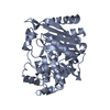
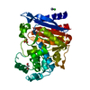
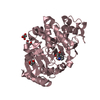
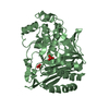
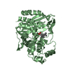
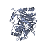
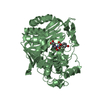
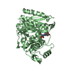
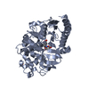
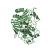
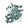
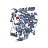
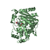
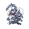
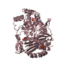
| Title | Crystal Structure of Beta-Lactamase/D-Alanine Carboxypeptidase from Pseudomonas syringae | ||||||
 Components Components | (Beta-Lactamase/D-Alanine ...) x 4 | ||||||
 Keywords Keywords | HYDROLASE / alpha-beta-fold / Structural Genomics / PSI-Biology / Midwest Center for Structural Genomics / MCSG | ||||||
| Function / homology |  Function and homology information Function and homology informationantibiotic catabolic process / beta-lactamase activity / beta-lactamase / outer membrane-bounded periplasmic space / response to antibiotic Similarity search - Function | ||||||
| Biological species |  Pseudomonas syringae pv. tomato (bacteria) Pseudomonas syringae pv. tomato (bacteria) | ||||||
| Method |  X-RAY DIFFRACTION / SYNCHROTRON / MOLECULAR REPLACEMENT / Resolution: 1.8 Å X-RAY DIFFRACTION / SYNCHROTRON / MOLECULAR REPLACEMENT / Resolution: 1.8 Å | ||||||
 Authors Authors | Kim, Y. / Hatzos-Skintges, C. / Endres, M. / Babnigg, G. / Joachimiak, A. / Midwest Center for Structural Genomics (MCSG) | ||||||
| Funding support |  United States, 1items United States, 1items
| ||||||
 Citation Citation | Journal: To Be Published Title: Crystal Structure of Beta-Lactamase/D-Alanine Carboxypeptidase from Pseudomonas syringae Authors: Kim, Y. / Hatzos-Skintges, C. / Endres, M. / Babnigg, G. / Joachimiak, A. / Midwest Center for Structural Genomics (MCSG) | ||||||
| History |
|
- Structure visualization
| Structure viewer | Molecule: MolmilJmol/JSmol |
|---|
- Downloads & links
-Download
| PDBx/mmCIF format | 5evi.cif.gz | 532.2 KB | Display | PDBx/mmCIF format |
|---|---|---|---|---|
| PDB format | pdb5evi.ent.gz | 438.3 KB | Display | PDB format |
| PDBx/mmJSON format | 5evi.json.gz | Tree view | PDBx/mmJSON format | |
| Others |  Other downloads Other downloads |
-Validation report
| Arichive directory | https://data.pdbj.org/pub/pdb/validation_reports/ev/5eviftp://data.pdbj.org/pub/pdb/validation_reports/ev/5evi | HTTPS FTP |
|---|
-Related structure data
| Similar structure data | |
|---|---|
| Other databases |


-Links
 PDBj
PDBj
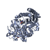
- Assembly
| Deposited unit | 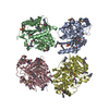
| ||||||||
|---|---|---|---|---|---|---|---|---|---|
| 1 | 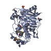
| ||||||||
| 2 | 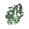
| ||||||||
| 3 | 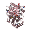
| ||||||||
| 4 | 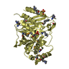
| ||||||||
| Unit cell |
|
-Components
-Beta-Lactamase/D-Alanine ... , 4 types, 4 molecules ABCD
| #1: Protein | Mass: 40686.734 Da / Num. of mol.: 1 Source method: isolated from a genetically manipulated source Source: (gene. exp.) Pseudomonas syringae pv. tomato (bacteria)Strain: DC3000 / Gene: ampC, PSPTO_3594 / Plasmid: pMCSG73 / Production host: |
|---|---|
| #2: Protein | Mass: 40686.730 Da / Num. of mol.: 1 Source method: isolated from a genetically manipulated source Source: (gene. exp.) Pseudomonas syringae pv. tomato (bacteria)Strain: DC3000 / Gene: ampC, PSPTO_3594 / Plasmid: pMCSG73 / Production host: |
| #3: Protein | Mass: 40686.734 Da / Num. of mol.: 1 Source method: isolated from a genetically manipulated source Source: (gene. exp.) Pseudomonas syringae pv. tomato (bacteria)Strain: DC3000 / Gene: ampC, PSPTO_3594 / Plasmid: pMCSG73 / Production host: |
| #4: Protein | Mass: 40713.777 Da / Num. of mol.: 1 Source method: isolated from a genetically manipulated source Source: (gene. exp.) Pseudomonas syringae pv. tomato (bacteria)Strain: DC3000 / Gene: ampC, PSPTO_3594 / Plasmid: pMCSG73 / Production host: |
-Non-polymers , 3 types, 523 molecules 


| #5: Chemical | ChemComp-SO4 /  Mass: 96.063 Da / Num. of mol.: 7 / Source method: obtained synthetically / Formula: SO4 Mass: 96.063 Da / Num. of mol.: 7 / Source method: obtained synthetically / Formula: SO4#6: Chemical | ChemComp-EDO /  Mass: 62.068 Da / Num. of mol.: 6 / Source method: obtained synthetically / Formula: C2H6O2 Mass: 62.068 Da / Num. of mol.: 6 / Source method: obtained synthetically / Formula: C2H6O2#7: Water | ChemComp-HOH / | Mass: 18.015 Da / Num. of mol.: 510 / Source method: isolated from a natural source / Formula: H2O |
|---|
-Details
| Has protein modification | Y |
|---|
-Experimental details
-Experiment
| Experiment | Method: X-RAY DIFFRACTION / Number of used crystals: 1 |
|---|
- Sample preparation
| Crystal | Density Matthews: 2.28 Å3/Da / Density % sol: 45.95 % |
|---|---|
| Crystal grow | Temperature: 289 K / Method: vapor diffusion, sitting drop / pH: 6.5 / Details: 2.0 M Ammonium Sulfate 0.1 M Bis-Tris:HCl pH 6.5 |
-Data collection
| Diffraction | Mean temperature: 100 K | |||||||||||||||||||||||||
|---|---|---|---|---|---|---|---|---|---|---|---|---|---|---|---|---|---|---|---|---|---|---|---|---|---|---|
| Diffraction source | Source: SYNCHROTRON / Site: APS / Beamline: 19-ID / Wavelength: 0.97926 Å | |||||||||||||||||||||||||
| Detector | Type: ADSC QUANTUM 315r / Detector: CCD / Date: Feb 8, 2015 | |||||||||||||||||||||||||
| Radiation | Monochromator: double crystal monochromator / Protocol: SINGLE WAVELENGTH / Monochromatic (M) / Laue (L): M / Scattering type: x-ray | |||||||||||||||||||||||||
| Radiation wavelength | Wavelength: 0.97926 Å / Relative weight: 1 | |||||||||||||||||||||||||
| Reflection twin |
| |||||||||||||||||||||||||
| Reflection | Resolution: 1.8→50 Å / Num. all: 128402 / Num. obs: 128402 / % possible obs: 97.5 % / Redundancy: 2.2 % / Rsym value: 0.054 / Net I/σ(I): 12.44 | |||||||||||||||||||||||||
| Reflection shell | Resolution: 1.8→1.83 Å / Redundancy: 2.1 % / Rmerge(I) obs: 0.419 / Mean I/σ(I) obs: 2.49 / % possible all: 96.2 |
- Processing
| Software |
| ||||||||||||||||||||||||||||||||||||||||||||||||||||||||||||||||||||||||||||||||||||||||||||||||||||||||||||||||||||||||||||||||||||||||||||||||||||||||||||||||||||||||||||||||||||||
|---|---|---|---|---|---|---|---|---|---|---|---|---|---|---|---|---|---|---|---|---|---|---|---|---|---|---|---|---|---|---|---|---|---|---|---|---|---|---|---|---|---|---|---|---|---|---|---|---|---|---|---|---|---|---|---|---|---|---|---|---|---|---|---|---|---|---|---|---|---|---|---|---|---|---|---|---|---|---|---|---|---|---|---|---|---|---|---|---|---|---|---|---|---|---|---|---|---|---|---|---|---|---|---|---|---|---|---|---|---|---|---|---|---|---|---|---|---|---|---|---|---|---|---|---|---|---|---|---|---|---|---|---|---|---|---|---|---|---|---|---|---|---|---|---|---|---|---|---|---|---|---|---|---|---|---|---|---|---|---|---|---|---|---|---|---|---|---|---|---|---|---|---|---|---|---|---|---|---|---|---|---|---|---|
| Refinement | Method to determine structure: MOLECULAR REPLACEMENT / Resolution: 1.8→36.32 Å / Cor.coef. Fo:Fc: 0.958 / Cor.coef. Fo:Fc free: 0.941 / SU B: 3.753 / SU ML: 0.064 / Cross valid method: THROUGHOUT / ESU R: 0.027 / ESU R Free: 0.025 / Stereochemistry target values: MAXIMUM LIKELIHOOD / Details: HYDROGENS HAVE BEEN ADDED IN THE RIDING POSITIONS
| ||||||||||||||||||||||||||||||||||||||||||||||||||||||||||||||||||||||||||||||||||||||||||||||||||||||||||||||||||||||||||||||||||||||||||||||||||||||||||||||||||||||||||||||||||||||
| Solvent computation | Ion probe radii: 0.8 Å / Shrinkage radii: 0.8 Å / VDW probe radii: 1.2 Å / Solvent model: MASK | ||||||||||||||||||||||||||||||||||||||||||||||||||||||||||||||||||||||||||||||||||||||||||||||||||||||||||||||||||||||||||||||||||||||||||||||||||||||||||||||||||||||||||||||||||||||
| Displacement parameters | Biso mean: 24.853 Å2
| ||||||||||||||||||||||||||||||||||||||||||||||||||||||||||||||||||||||||||||||||||||||||||||||||||||||||||||||||||||||||||||||||||||||||||||||||||||||||||||||||||||||||||||||||||||||
| Refinement step | Cycle: LAST / Resolution: 1.8→36.32 Å
| ||||||||||||||||||||||||||||||||||||||||||||||||||||||||||||||||||||||||||||||||||||||||||||||||||||||||||||||||||||||||||||||||||||||||||||||||||||||||||||||||||||||||||||||||||||||
| Refine LS restraints |
|