Protocol: SINGLE WAVELENGTH / Monochromatic (M) / Laue (L): M / Scattering type: x-ray
Radiation wavelength
Wavelength: 1.1 Å / Relative weight: 1
Reflection
Redundancy: 3.5 % / Av σ(I) over netI: 5.6 / Number: 182821 / Rmerge(I) obs: 0.126 / Χ2: 1.59 / D res high: 2 Å / D res low: 50 Å / Num. obs: 52485 / % possible obs: 92.2
Diffraction reflection shell
Highest resolution (Å)
Lowest resolution (Å)
% possible obs (%)
ID
Rmerge(I) obs
Chi squared
Redundancy
4.31
50
99.9
1
0.073
2.545
3.8
3.42
4.31
100
1
0.083
1.972
4
2.99
3.42
99.9
1
0.12
1.583
3.8
2.71
2.99
98.9
1
0.189
1.313
3.7
2.52
2.71
96.5
1
0.28
1.272
3.7
2.37
2.52
94
1
0.342
1.273
3.6
2.25
2.37
92.4
1
0.388
1.506
3.3
2.15
2.25
88.3
1
0.408
1.245
3.1
2.07
2.15
80.4
1
0.448
1.182
2.9
2
2.07
71
1
0.5
1.272
2.5
Reflection
Resolution: 2→50 Å / Num. obs: 52485 / % possible obs: 92.2 % / Redundancy: 3.5 % / Rmerge(I) obs: 0.126 / Χ2: 1.585 / Net I/σ(I): 5.6
Reflection shell
Resolution (Å)
Redundancy (%)
Rmerge(I) obs
Num. unique all
Χ2
Diffraction-ID
% possible all
2-2.07
2.5
0.5
4001
1.272
1
71
2.07-2.15
2.9
0.448
4498
1.182
1
80.4
2.15-2.25
3.1
0.408
4955
1.245
1
88.3
2.25-2.37
3.3
0.388
5186
1.506
1
92.4
2.37-2.52
3.6
0.342
5313
1.273
1
94
2.52-2.71
3.7
0.28
5448
1.272
1
96.5
2.71-2.99
3.7
0.189
5632
1.313
1
98.9
2.99-3.42
3.8
0.12
5698
1.583
1
99.9
3.42-4.31
4
0.083
5752
1.972
1
100
4.31-50
3.8
0.073
6002
2.545
1
99.9
-
Phasing
Phasing MR
Highest resolution
Lowest resolution
Rotation
2.5 Å
45.88 Å
Translation
2.5 Å
45.88 Å
-
Processing
Software
Name
Version
Classification
NB
DENZO
datareduction
SCALEPACK
datascaling
PHASER
phasing
REFMAC
5.2.0019
refinement
PDB_EXTRACT
2
dataextraction
Refinement
Method to determine structure: MOLECULAR REPLACEMENT / Resolution: 2→30 Å / Cor.coef. Fo:Fc: 0.956 / Cor.coef. Fo:Fc free: 0.93 / SU B: 9.87 / SU ML: 0.124 / TLS residual ADP flag: LIKELY RESIDUAL / Cross valid method: THROUGHOUT / σ(F): 0 / ESU R: 0.205 / ESU R Free: 0.179 / Stereochemistry target values: MAXIMUM LIKELIHOOD
Rfactor
Num. reflection
% reflection
Selection details
Rfree
0.239
2642
5.1 %
RANDOM
Rwork
0.193
-
-
-
obs
0.195
52067
91.95 %
-
Solvent computation
Ion probe radii: 0.8 Å / Shrinkage radii: 0.8 Å / VDW probe radii: 1.2 Å / Solvent model: MASK
Displacement parameters
Biso mean: 26.489 Å2
Baniso -1
Baniso -2
Baniso -3
1-
2.41 Å2
0 Å2
0 Å2
2-
-
-0.7 Å2
0 Å2
3-
-
-
-1.71 Å2
Refinement step
Cycle: LAST / Resolution: 2→30 Å
Protein
Nucleic acid
Ligand
Solvent
Total
Num. atoms
5600
0
52
621
6273
Refine LS restraints
Refine-ID
Type
Dev ideal
Dev ideal target
Number
X-RAY DIFFRACTION
r_bond_refined_d
0.013
0.022
5808
X-RAY DIFFRACTION
r_angle_refined_deg
1.469
1.956
7936
X-RAY DIFFRACTION
r_dihedral_angle_1_deg
6.408
5
714
X-RAY DIFFRACTION
r_dihedral_angle_2_deg
34.805
24.797
246
X-RAY DIFFRACTION
r_dihedral_angle_3_deg
14.539
15
912
X-RAY DIFFRACTION
r_dihedral_angle_4_deg
13.388
15
22
X-RAY DIFFRACTION
r_chiral_restr
0.089
0.2
856
X-RAY DIFFRACTION
r_gen_planes_refined
0.005
0.02
4450
X-RAY DIFFRACTION
r_nbd_refined
0.21
0.2
2629
X-RAY DIFFRACTION
r_nbtor_refined
0.301
0.2
3909
X-RAY DIFFRACTION
r_xyhbond_nbd_refined
0.155
0.2
503
X-RAY DIFFRACTION
r_symmetry_vdw_refined
0.217
0.2
64
X-RAY DIFFRACTION
r_symmetry_hbond_refined
0.186
0.2
24
X-RAY DIFFRACTION
r_mcbond_it
0.686
1.5
3676
X-RAY DIFFRACTION
r_mcangle_it
1.078
2
5772
X-RAY DIFFRACTION
r_scbond_it
1.792
3
2529
X-RAY DIFFRACTION
r_scangle_it
2.706
4.5
2164
LS refinement shell
Resolution: 2.003→2.055 Å / Total num. of bins used: 20
Rfactor
Num. reflection
% reflection
Rfree
0.3
122
-
Rwork
0.236
2573
-
obs
-
2695
65.67 %
Refinement TLS params.
Method: refined / Refine-ID: X-RAY DIFFRACTION
ID
L11 (°2)
L12 (°2)
L13 (°2)
L22 (°2)
L23 (°2)
L33 (°2)
S11 (Å °)
S12 (Å °)
S13 (Å °)
S21 (Å °)
S22 (Å °)
S23 (Å °)
S31 (Å °)
S32 (Å °)
S33 (Å °)
T11 (Å2)
T12 (Å2)
T13 (Å2)
T22 (Å2)
T23 (Å2)
T33 (Å2)
Origin x (Å)
Origin y (Å)
Origin z (Å)
1
0.5973
0.2824
0.3274
0.3434
0.1775
0.6456
-0.039
-0.0545
0.0222
-0.0564
-0.0282
-0.0149
0.0383
-0.0444
0.0672
-0.0005
0.0053
0.0172
-0.0529
0.0009
-0.0511
34.6431
-2.6715
-8.9287
2
0.2376
-0.1371
-0.258
0.4746
0.1949
0.8258
0.0211
0.0151
-0.0075
0.0288
-0.042
-0.0252
-0.0131
-0.0063
0.0209
-0.0074
-0.0071
-0.0093
-0.0376
0.0057
-0.0472
35.389
32.9269
-32.5586
3
0.0359
0.005
0.0035
0.041
-0.0201
0.0564
-0.0158
0.0036
0.0016
-0.0042
-0.008
-0.0122
-0.0036
-0.0157
0.0237
0.0298
0.0001
0.0036
0.0173
0.0027
0.0212
34.6815
14.467
-20.2761
Refinement TLS group
ID
Refine-ID
Refine TLS-ID
Auth asym-ID
Auth seq-ID
1
X-RAY DIFFRACTION
1
A
4 - 361
2
X-RAY DIFFRACTION
2
B
4 - 361
3
X-RAY DIFFRACTION
3
A
803
4
X-RAY DIFFRACTION
3
B
701 - 804
5
X-RAY DIFFRACTION
3
A
902 - 1235
6
X-RAY DIFFRACTION
3
B
903 - 1189
+
About Yorodumi
-
News
-
Feb 9, 2022. New format data for meta-information of EMDB entries
New format data for meta-information of EMDB entries
Version 3 of the EMDB header file is now the official format.
The previous official version 1.9 will be removed from the archive.
In the structure databanks used in Yorodumi, some data are registered as the other names, "COVID-19 virus" and "2019-nCoV". Here are the details of the virus and the list of structure data.
Jan 31, 2019. EMDB accession codes are about to change! (news from PDBe EMDB page)
EMDB accession codes are about to change! (news from PDBe EMDB page)
The allocation of 4 digits for EMDB accession codes will soon come to an end. Whilst these codes will remain in use, new EMDB accession codes will include an additional digit and will expand incrementally as the available range of codes is exhausted. The current 4-digit format prefixed with “EMD-” (i.e. EMD-XXXX) will advance to a 5-digit format (i.e. EMD-XXXXX), and so on. It is currently estimated that the 4-digit codes will be depleted around Spring 2019, at which point the 5-digit format will come into force.
The EM Navigator/Yorodumi systems omit the EMD- prefix.
Related info.:Q: What is EMD? / ID/Accession-code notation in Yorodumi/EM Navigator
Yorodumi is a browser for structure data from EMDB, PDB, SASBDB, etc.
This page is also the successor to EM Navigator detail page, and also detail information page/front-end page for Omokage search.
The word "yorodu" (or yorozu) is an old Japanese word meaning "ten thousand". "mi" (miru) is to see.
Related info.:EMDB / PDB / SASBDB / Comparison of 3 databanks / Yorodumi Search / Aug 31, 2016. New EM Navigator & Yorodumi / Yorodumi Papers / Jmol/JSmol / Function and homology information / Changes in new EM Navigator and Yorodumi
 Movie
Movie Controller


 Open data
 Basic information
 Components
 Keywords
 Function and homology information
 X-RAY DIFFRACTION /
  Authors
 Citation
 Structure visualization
 Downloads & links
 Other downloads


 PDBj
PDBj
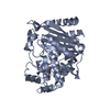
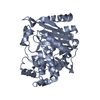
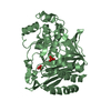
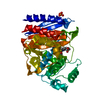
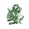
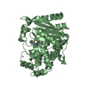
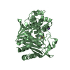
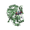
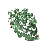
 Assembly


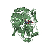
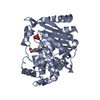
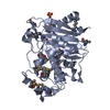
 Mass: 78.133 Da / Num. of mol.: 2 / Source method: obtained synthetically / Formula: C2H6OS / Comment: DMSO, precipitant*YM
 Mass: 241.287 Da / Num. of mol.: 1 / Source method: obtained synthetically / Formula: C11H19N3O3
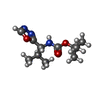
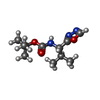
 Mass: 96.063 Da / Num. of mol.: 2 / Source method: obtained synthetically / Formula: SO4
 Mass: 241.287 Da / Num. of mol.: 1 / Source method: obtained synthetically / Formula: C11H19N3O3
 Sample preparation
 / Beamline: 8.3.1 / Wavelength: 1.1
  Processing