 Movie
Movie Controller

[English] 日本語
 Yorodumi
- PDB-1pi5: Structure of N289A mutant of AmpC in complex with SM2, carboxyphe... -
+ Open data

- Basic information
| Entry | Database: PDB / ID: 1pi5 | ||||||
|---|---|---|---|---|---|---|---|
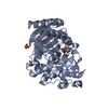
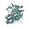
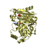
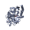
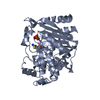
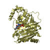
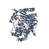
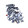
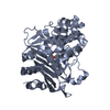
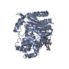
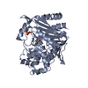
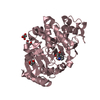
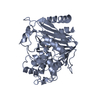
| Title | Structure of N289A mutant of AmpC in complex with SM2, carboxyphenylglycylboronic acid bearing the cephalothin R1 side chain | ||||||
 Components Components | Beta-lactamase | ||||||
 Keywords Keywords | HYDROLASE / Enzyme Inhibitor Complex / Beta-Lactamase | ||||||
| Function / homology |  Function and homology information Function and homology informationantibiotic catabolic process / beta-lactamase activity / beta-lactamase / outer membrane-bounded periplasmic space / response to antibiotic Similarity search - Function | ||||||
| Biological species |  | ||||||
| Method |  X-RAY DIFFRACTION / SYNCHROTRON / MOLECULAR REPLACEMENT / Resolution: 1.49 Å X-RAY DIFFRACTION / SYNCHROTRON / MOLECULAR REPLACEMENT / Resolution: 1.49 Å | ||||||
 Authors Authors | Roth, T.A. / Minasov, G. / Focia, P.J. / Shoichet, B.K. | ||||||
 Citation Citation | Journal: Biochemistry / Year: 2003 Title: Thermodynamic cycle analysis and inhibitor design against beta-lactamase. Authors: Roth, T.A. / Minasov, G. / Morandi, S. / Prati, F. / Shoichet, B.K. | ||||||
| History |
|
- Structure visualization
| Structure viewer | Molecule: MolmilJmol/JSmol |
|---|
- Downloads & links
-Download
| PDBx/mmCIF format | 1pi5.cif.gz | 192.6 KB | Display | PDBx/mmCIF format |
|---|---|---|---|---|
| PDB format | pdb1pi5.ent.gz | 152.8 KB | Display | PDB format |
| PDBx/mmJSON format | 1pi5.json.gz | Tree view | PDBx/mmJSON format | |
| Others |  Other downloads Other downloads |
-Validation report
| Arichive directory | https://data.pdbj.org/pub/pdb/validation_reports/pi/1pi5ftp://data.pdbj.org/pub/pdb/validation_reports/pi/1pi5 | HTTPS FTP |
|---|
-Related structure data
| Related structure data | 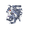 1pi4C 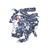 1mxoS S: Starting model for refinement C: citing same article ( |
|---|---|
| Similar structure data |


-Links
 PDBj
PDBj
- Assembly
| Deposited unit | 
| ||||||||
|---|---|---|---|---|---|---|---|---|---|
| 1 | 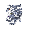
| ||||||||
| 2 | 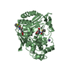
| ||||||||
| Unit cell |
|
-Components
| #1: Protein | Mass: 39544.898 Da / Num. of mol.: 2 / Fragment: AmpC / Mutation: N289A Source method: isolated from a genetically manipulated source Source: (gene. exp.) #2: Chemical | ChemComp-PO4 / |   Mass: 94.971 Da / Num. of mol.: 1 / Source method: obtained synthetically / Formula: PO4 Mass: 94.971 Da / Num. of mol.: 1 / Source method: obtained synthetically / Formula: PO4#3: Chemical |   Mass: 39.098 Da / Num. of mol.: 2 / Source method: obtained synthetically / Formula: K Mass: 39.098 Da / Num. of mol.: 2 / Source method: obtained synthetically / Formula: K#4: Chemical |   Mass: 319.141 Da / Num. of mol.: 3 / Source method: obtained synthetically / Formula: C14H14BNO5S Mass: 319.141 Da / Num. of mol.: 3 / Source method: obtained synthetically / Formula: C14H14BNO5S#5: Water | ChemComp-HOH / |  Mass: 18.015 Da / Num. of mol.: 1179 / Source method: isolated from a natural source / Formula: H2O Mass: 18.015 Da / Num. of mol.: 1179 / Source method: isolated from a natural source / Formula: H2OHas protein modification | Y | |
|---|
-Experimental details
-Experiment
| Experiment | Method: X-RAY DIFFRACTION / Number of used crystals: 1 |
|---|
- Sample preparation
| Crystal | Density Matthews: 1.91 Å3/Da / Density % sol: 35.05 % |
|---|---|
| Crystal grow | Temperature: 295 K / Method: vapor diffusion, hanging drop / pH: 8.7 Details: Potassium Phosphate Buffer , pH 8.7, VAPOR DIFFUSION, HANGING DROP, temperature 295K |
-Data collection
| Diffraction | Mean temperature: 100 K |
|---|---|
| Diffraction source | Source: SYNCHROTRON / Site: APS  / Beamline: 5ID-B / Wavelength: 1 Å / Beamline: 5ID-B / Wavelength: 1 Å |
| Detector | Type: MARRESEARCH / Detector: CCD / Date: Dec 1, 2002 / Details: Mirrors |
| Radiation | Monochromator: silicon / Protocol: SINGLE WAVELENGTH / Monochromatic (M) / Laue (L): M / Scattering type: x-ray |
| Radiation wavelength | Wavelength: 1 Å / Relative weight: 1 |
| Reflection | Resolution: 1.49→15 Å / Num. all: 128092 / Num. obs: 128092 / % possible obs: 99.9 % / Observed criterion σ(I): -3 / Redundancy: 3.8 % / Biso Wilson estimate: 22.8 Å2 / Rmerge(I) obs: 0.051 / Net I/σ(I): 23.8 |
| Reflection shell | Resolution: 1.49→1.54 Å / Redundancy: 3.4 % / Rmerge(I) obs: 0.184 / Mean I/σ(I) obs: 7.6 / Num. unique all: 12767 / % possible all: 100 |
| Reflection | *PLUS Num. measured all: 483237 |
| Reflection shell | *PLUS % possible obs: 100 % / Num. unique obs: 12767 |
- Processing
| Software |
| ||||||||||||||||||||||||||||||||||||||||||||||||||||||||||||||||||||||||||||||||
|---|---|---|---|---|---|---|---|---|---|---|---|---|---|---|---|---|---|---|---|---|---|---|---|---|---|---|---|---|---|---|---|---|---|---|---|---|---|---|---|---|---|---|---|---|---|---|---|---|---|---|---|---|---|---|---|---|---|---|---|---|---|---|---|---|---|---|---|---|---|---|---|---|---|---|---|---|---|---|---|---|---|
| Refinement | Method to determine structure: MOLECULAR REPLACEMENT Starting model: 1MXO Resolution: 1.49→15 Å Isotropic thermal model: Isotropic with refined individual B-factors Cross valid method: THROUGHOUT / σ(F): 0 / σ(I): 0 / Stereochemistry target values: Engh & Huber / Details: Maximum Likelihood in the refinement
| ||||||||||||||||||||||||||||||||||||||||||||||||||||||||||||||||||||||||||||||||
| Solvent computation | Bsol: 250 Å2 / ksol: 0.647 e/Å3 | ||||||||||||||||||||||||||||||||||||||||||||||||||||||||||||||||||||||||||||||||
| Displacement parameters | Biso mean: 18.2 Å2
| ||||||||||||||||||||||||||||||||||||||||||||||||||||||||||||||||||||||||||||||||
| Refine analyze |
| ||||||||||||||||||||||||||||||||||||||||||||||||||||||||||||||||||||||||||||||||
| Refinement step | Cycle: LAST / Resolution: 1.49→15 Å
| ||||||||||||||||||||||||||||||||||||||||||||||||||||||||||||||||||||||||||||||||
| Refine LS restraints |
| ||||||||||||||||||||||||||||||||||||||||||||||||||||||||||||||||||||||||||||||||
| LS refinement shell | Resolution: 1.49→1.54 Å
| ||||||||||||||||||||||||||||||||||||||||||||||||||||||||||||||||||||||||||||||||
| Refinement | *PLUS Rfactor Rwork: 0.156 | ||||||||||||||||||||||||||||||||||||||||||||||||||||||||||||||||||||||||||||||||
| Solvent computation | *PLUS | ||||||||||||||||||||||||||||||||||||||||||||||||||||||||||||||||||||||||||||||||
| Displacement parameters | *PLUS | ||||||||||||||||||||||||||||||||||||||||||||||||||||||||||||||||||||||||||||||||
| Refine LS restraints | *PLUS
|
