 Movie
Movie Controller

[English] 日本語
 Yorodumi
- PDB-1iem: Crystal Structure of AmpC beta-lactamase from E. coli in Complex ... -
+ Open data

- Basic information
| Entry | Database: PDB / ID: 1iem | ||||||
|---|---|---|---|---|---|---|---|
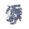
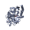
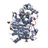
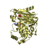
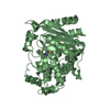
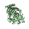
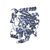
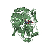
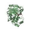
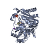
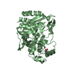
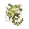
| Title | Crystal Structure of AmpC beta-lactamase from E. coli in Complex with a Boronic Acid Inhibitor (1, CefB4) | ||||||
 Components Components | beta-lactamase | ||||||
 Keywords Keywords | HYDROLASE / cephalosporinase / beta-lactamase / serine hydrolase | ||||||
| Function / homology |  Function and homology information Function and homology informationantibiotic catabolic process / beta-lactamase activity / beta-lactamase / outer membrane-bounded periplasmic space / response to antibiotic Similarity search - Function | ||||||
| Biological species |  | ||||||
| Method |  X-RAY DIFFRACTION / SYNCHROTRON / MOLECULAR REPLACEMENT / Resolution: 2.3 Å X-RAY DIFFRACTION / SYNCHROTRON / MOLECULAR REPLACEMENT / Resolution: 2.3 Å | ||||||
 Authors Authors | Powers, R.A. / Caselli, E. / Focia, P.J. / Prati, F. / Shoichet, B.K. | ||||||
 Citation Citation | Journal: Biochemistry / Year: 2001 Title: Structures of ceftazidime and its transition-state analogue in complex with AmpC beta-lactamase: implications for resistance mutations and inhibitor design. Authors: Powers, R.A. / Caselli, E. / Focia, P.J. / Prati, F. / Shoichet, B.K. | ||||||
| History |
|
- Structure visualization
| Structure viewer | Molecule: MolmilJmol/JSmol |
|---|
- Downloads & links
-Download
| PDBx/mmCIF format | 1iem.cif.gz | 152.7 KB | Display | PDBx/mmCIF format |
|---|---|---|---|---|
| PDB format | pdb1iem.ent.gz | 120.2 KB | Display | PDB format |
| PDBx/mmJSON format | 1iem.json.gz | Tree view | PDBx/mmJSON format | |
| Others |  Other downloads Other downloads |
-Validation report
| Arichive directory | https://data.pdbj.org/pub/pdb/validation_reports/ie/1iemftp://data.pdbj.org/pub/pdb/validation_reports/ie/1iem | HTTPS FTP |
|---|
-Related structure data
| Related structure data |  1ielC  1fsyS S: Starting model for refinement C: citing same article ( |
|---|---|
| Similar structure data |


-Links
 PDBj
PDBj
- Assembly
| Deposited unit | 
| ||||||||
|---|---|---|---|---|---|---|---|---|---|
| 1 | 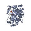
| ||||||||
| 2 | 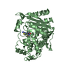
| ||||||||
| Unit cell |
|
-Components
| #1: Protein | Mass: 39587.922 Da / Num. of mol.: 2 Source method: isolated from a genetically manipulated source Source: (gene. exp.) #2: Chemical | ChemComp-PO4 / |   Mass: 94.971 Da / Num. of mol.: 1 / Source method: obtained synthetically / Formula: PO4 Mass: 94.971 Da / Num. of mol.: 1 / Source method: obtained synthetically / Formula: PO4#3: Chemical |   Mass: 330.125 Da / Num. of mol.: 2 / Source method: obtained synthetically / Formula: C10H15BN4O6S Mass: 330.125 Da / Num. of mol.: 2 / Source method: obtained synthetically / Formula: C10H15BN4O6S#4: Water | ChemComp-HOH / |  Mass: 18.015 Da / Num. of mol.: 209 / Source method: isolated from a natural source / Formula: H2O Mass: 18.015 Da / Num. of mol.: 209 / Source method: isolated from a natural source / Formula: H2OHas protein modification | Y | |
|---|
-Experimental details
-Experiment
| Experiment | Method: X-RAY DIFFRACTION / Number of used crystals: 1 |
|---|
- Sample preparation
| Crystal | Density Matthews: 2.56 Å3/Da / Density % sol: 52.03 % | |||||||||||||||||||||||||
|---|---|---|---|---|---|---|---|---|---|---|---|---|---|---|---|---|---|---|---|---|---|---|---|---|---|---|
| Crystal grow | Temperature: 296 K / Method: vapor diffusion, hanging drop / pH: 8.7 Details: 1.7M potassium phosphate, 95uM AmpC, 586uM compound 1 (CB4), pH 8.7, VAPOR DIFFUSION, HANGING DROP, temperature 296K | |||||||||||||||||||||||||
| Crystal grow | *PLUS Temperature: 23 ℃ / Details: used microseeding | |||||||||||||||||||||||||
| Components of the solutions | *PLUS
|
-Data collection
| Diffraction | Mean temperature: 100 K |
|---|---|
| Diffraction source | Source: SYNCHROTRON / Site: SSRL  / Beamline: BL7-1 / Wavelength: 1.08 Å / Beamline: BL7-1 / Wavelength: 1.08 Å |
| Detector | Type: MARRESEARCH / Detector: IMAGE PLATE / Date: Jul 4, 2000 |
| Radiation | Protocol: SINGLE WAVELENGTH / Monochromatic (M) / Laue (L): M / Scattering type: x-ray |
| Radiation wavelength | Wavelength: 1.08 Å / Relative weight: 1 |
| Reflection | Resolution: 2.3→20 Å / Num. all: 35024 / Num. obs: 137203 / % possible obs: 98.8 % / Observed criterion σ(I): -3 / Redundancy: 3.9 % / Biso Wilson estimate: 26.68 Å2 / Rmerge(I) obs: 0.075 / Net I/σ(I): 11.2 |
| Reflection shell | Resolution: 2.3→2.38 Å / Rmerge(I) obs: 0.359 / % possible all: 99.9 |
| Reflection | *PLUS Lowest resolution: 20 Å / Num. obs: 35024 / Num. measured all: 137203 |
| Reflection shell | *PLUS % possible obs: 99.9 % / Mean I/σ(I) obs: 4 |
- Processing
| Software |
| |||||||||||||||||||||||||
|---|---|---|---|---|---|---|---|---|---|---|---|---|---|---|---|---|---|---|---|---|---|---|---|---|---|---|
| Refinement | Method to determine structure: MOLECULAR REPLACEMENT Starting model: PDB entry 1FSY Resolution: 2.3→20 Å / σ(F): 2 / Stereochemistry target values: Engh & Huber
| |||||||||||||||||||||||||
| Refine analyze | Luzzati coordinate error obs: 0.25 Å / Luzzati d res low obs: 5 Å | |||||||||||||||||||||||||
| Refinement step | Cycle: LAST / Resolution: 2.3→20 Å
| |||||||||||||||||||||||||
| Refine LS restraints |
| |||||||||||||||||||||||||
| LS refinement shell | Resolution: 2.3→2.4 Å
| |||||||||||||||||||||||||
| Software | *PLUS Name: CNS / Version: 1 / Classification: refinement | |||||||||||||||||||||||||
| Refinement | *PLUS Lowest resolution: 20 Å / σ(F): 2 / % reflection Rfree: 6 % / Rfactor obs: 0.182 | |||||||||||||||||||||||||
| Solvent computation | *PLUS | |||||||||||||||||||||||||
| Displacement parameters | *PLUS | |||||||||||||||||||||||||
| LS refinement shell | *PLUS Highest resolution: 2.3 Å / Lowest resolution: 2.4 Å / Rfactor Rfree: 0.278 / Rfactor Rwork: 0.217 |
