 Movie
Movie Controller

[English] 日本語
 Yorodumi
- PDB-4kg5: Crystal Structure of AmpC beta-lactamase N152G Mutant in Complex ... -
+ Open data

- Basic information
| Entry | Database: PDB / ID: 4kg5 | |||||||||
|---|---|---|---|---|---|---|---|---|---|---|
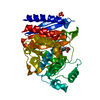
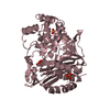
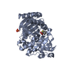
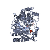
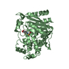
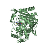
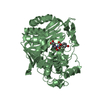
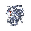
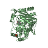
| Title | Crystal Structure of AmpC beta-lactamase N152G Mutant in Complex with Cefotaxime | |||||||||
 Components Components | Beta-lactamase | |||||||||
 Keywords Keywords | Hydrolase/antibiotic / Cephalosporinase / beta-lactamase / serine hydrolase / acyl-enzyme complex / Hydrolase-antibiotic complex | |||||||||
| Function / homology |  Function and homology information Function and homology informationantibiotic catabolic process / beta-lactamase / beta-lactamase activity / outer membrane-bounded periplasmic space / response to antibiotic Similarity search - Function | |||||||||
| Biological species |  | |||||||||
| Method |  X-RAY DIFFRACTION / SYNCHROTRON / MOLECULAR REPLACEMENT / Resolution: 2.11 Å X-RAY DIFFRACTION / SYNCHROTRON / MOLECULAR REPLACEMENT / Resolution: 2.11 Å | |||||||||
 Authors Authors | Docter, B.E. / Baggett, V.L. / Powers, R.A. / Wallar, B.J. | |||||||||
 Citation Citation | Journal: To be Published Title: Complexed structures of AmpC beta-lactamase Authors: Docter, B.E. / Baggett, V.L. / Powers, R.A. / Wallar, B.J. | |||||||||
| History |
|
- Structure visualization
| Structure viewer | Molecule: MolmilJmol/JSmol |
|---|
- Downloads & links
-Download
| PDBx/mmCIF format | 4kg5.cif.gz | 293.7 KB | Display | PDBx/mmCIF format |
|---|---|---|---|---|
| PDB format | pdb4kg5.ent.gz | 238.7 KB | Display | PDB format |
| PDBx/mmJSON format | 4kg5.json.gz | Tree view | PDBx/mmJSON format | |
| Others |  Other downloads Other downloads |
-Validation report
| Arichive directory | https://data.pdbj.org/pub/pdb/validation_reports/kg/4kg5ftp://data.pdbj.org/pub/pdb/validation_reports/kg/4kg5 | HTTPS FTP |
|---|
-Related structure data


-Links
 PDBj
PDBj
- Assembly
| Deposited unit | 
| ||||||||
|---|---|---|---|---|---|---|---|---|---|
| 1 | 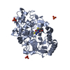
| ||||||||
| 2 | 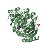
| ||||||||
| 3 | 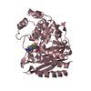
| ||||||||
| 4 | 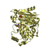
| ||||||||
| Unit cell |
|
-Components
| #1: Protein | Mass: 39530.871 Da / Num. of mol.: 4 / Mutation: N152G Source method: isolated from a genetically manipulated source Source: (gene. exp.) #2: Chemical | ChemComp-PO4 /   Mass: 94.971 Da / Num. of mol.: 5 / Source method: obtained synthetically / Formula: PO4 Mass: 94.971 Da / Num. of mol.: 5 / Source method: obtained synthetically / Formula: PO4#3: Chemical | ChemComp-CEF / 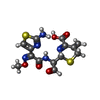  Mass: 397.429 Da / Num. of mol.: 4 / Source method: obtained synthetically / Formula: C14H15N5O5S2 Mass: 397.429 Da / Num. of mol.: 4 / Source method: obtained synthetically / Formula: C14H15N5O5S2#4: Water | ChemComp-HOH / |  Mass: 18.015 Da / Num. of mol.: 496 / Source method: isolated from a natural source / Formula: H2O Mass: 18.015 Da / Num. of mol.: 496 / Source method: isolated from a natural source / Formula: H2OHas protein modification | Y | |
|---|
-Experimental details
-Experiment
| Experiment | Method: X-RAY DIFFRACTION / Number of used crystals: 1 |
|---|
- Sample preparation
| Crystal | Density Matthews: 2.61 Å3/Da / Density % sol: 52.94 % |
|---|---|
| Crystal grow | Temperature: 296 K / Method: vapor diffusion, hanging drop / pH: 8.7 Details: 1.7M potassium phosphate, 3.5mg/ml AmpC, pH 8.7, VAPOR DIFFUSION, HANGING DROP, temperature 296K |
-Data collection
| Diffraction | Mean temperature: 100 K |
|---|---|
| Diffraction source | Source: SYNCHROTRON / Site: APS  / Beamline: 21-ID-D / Wavelength: 1.1 Å / Beamline: 21-ID-D / Wavelength: 1.1 Å |
| Detector | Type: MAR scanner 300 mm plate / Detector: IMAGE PLATE / Date: Mar 10, 2012 |
| Radiation | Protocol: SINGLE WAVELENGTH / Monochromatic (M) / Laue (L): M / Scattering type: x-ray |
| Radiation wavelength | Wavelength: 1.1 Å / Relative weight: 1 |
| Reflection | Resolution: 2.11→50 Å / Num. obs: 91517 |
| Reflection shell | Resolution: 2.11→2.18 Å / % possible all: 99.9 |
- Processing
| Software |
| ||||||||||||||||||||||||||||||||||||||||||||||||||||||||||||||||||||||||||||||||||||||||||||||||||||||||||||||||||||||||||||||||||||||||||||||||||||||||||||||||||||||||||
|---|---|---|---|---|---|---|---|---|---|---|---|---|---|---|---|---|---|---|---|---|---|---|---|---|---|---|---|---|---|---|---|---|---|---|---|---|---|---|---|---|---|---|---|---|---|---|---|---|---|---|---|---|---|---|---|---|---|---|---|---|---|---|---|---|---|---|---|---|---|---|---|---|---|---|---|---|---|---|---|---|---|---|---|---|---|---|---|---|---|---|---|---|---|---|---|---|---|---|---|---|---|---|---|---|---|---|---|---|---|---|---|---|---|---|---|---|---|---|---|---|---|---|---|---|---|---|---|---|---|---|---|---|---|---|---|---|---|---|---|---|---|---|---|---|---|---|---|---|---|---|---|---|---|---|---|---|---|---|---|---|---|---|---|---|---|---|---|---|---|---|---|
| Refinement | Method to determine structure: MOLECULAR REPLACEMENT / Resolution: 2.11→28.23 Å / Cor.coef. Fo:Fc: 0.968 / Cor.coef. Fo:Fc free: 0.946 / SU B: 5.155 / SU ML: 0.133 / Cross valid method: THROUGHOUT / ESU R: 0.211 / ESU R Free: 0.185 / Stereochemistry target values: MAXIMUM LIKELIHOOD / Details: HYDROGENS HAVE BEEN ADDED IN THE RIDING POSITIONS
| ||||||||||||||||||||||||||||||||||||||||||||||||||||||||||||||||||||||||||||||||||||||||||||||||||||||||||||||||||||||||||||||||||||||||||||||||||||||||||||||||||||||||||
| Solvent computation | Ion probe radii: 0.8 Å / Shrinkage radii: 0.8 Å / VDW probe radii: 1.2 Å / Solvent model: BABINET MODEL WITH MASK | ||||||||||||||||||||||||||||||||||||||||||||||||||||||||||||||||||||||||||||||||||||||||||||||||||||||||||||||||||||||||||||||||||||||||||||||||||||||||||||||||||||||||||
| Displacement parameters | Biso mean: 36.593 Å2
| ||||||||||||||||||||||||||||||||||||||||||||||||||||||||||||||||||||||||||||||||||||||||||||||||||||||||||||||||||||||||||||||||||||||||||||||||||||||||||||||||||||||||||
| Refinement step | Cycle: LAST / Resolution: 2.11→28.23 Å
| ||||||||||||||||||||||||||||||||||||||||||||||||||||||||||||||||||||||||||||||||||||||||||||||||||||||||||||||||||||||||||||||||||||||||||||||||||||||||||||||||||||||||||
| Refine LS restraints |
| ||||||||||||||||||||||||||||||||||||||||||||||||||||||||||||||||||||||||||||||||||||||||||||||||||||||||||||||||||||||||||||||||||||||||||||||||||||||||||||||||||||||||||
| LS refinement shell | Resolution: 2.11→2.162 Å / Total num. of bins used: 20
|
