[English] 日本語
Yorodumi
- PDB-6o8g: Crystal structure of UvrB bound to fully duplex DNA -

+
Open data


ID or keywords:

Loading...

-
Basic information

Entry
Database: PDB / ID: 6o8g
TitleCrystal structure of UvrB bound to fully duplex DNA
Components
  • DNA (5'-D(*GP*GP*TP*AP*GP*CP*GP*CP*GP*AP*TP*GP*GP*AP*GP*A)-3')
  • DNA (5'-D(*TP*CP*TP*CP*CP*AP*TP*CP*GP*CP*GP*CP*TP*AP*C)-3')
  • UvrABC system protein B
KeywordsDNA BINDING PROTEIN/DNA / DNA repair / nucleotide excision repair / DNA BINDING PROTEIN-DNA complex
Function / homology
Function and homology information


excinuclease ABC activity / excinuclease repair complex / SOS response / nucleotide-excision repair / ATP hydrolysis activity / DNA binding / ATP binding / cytoplasm
Similarity search - Function
UvrB, YAD/RRR-motif-containing domain / Ultra-violet resistance protein B / UvrABC system, subunit B / UVR domain superfamily / UvrB/uvrC motif / UvrB, interaction domain / UvrB interaction domain / UVR domain / UVR domain profile. / Helicase/UvrB, N-terminal ...UvrB, YAD/RRR-motif-containing domain / Ultra-violet resistance protein B / UvrABC system, subunit B / UVR domain superfamily / UvrB/uvrC motif / UvrB, interaction domain / UvrB interaction domain / UVR domain / UVR domain profile. / Helicase/UvrB, N-terminal / Type III restriction enzyme, res subunit / Helicase conserved C-terminal domain / helicase superfamily c-terminal domain / Superfamilies 1 and 2 helicase C-terminal domain profile. / Superfamilies 1 and 2 helicase ATP-binding type-1 domain profile. / DEAD-like helicases superfamily / Helicase, C-terminal / Helicase superfamily 1/2, ATP-binding domain / P-loop containing nucleoside triphosphate hydrolase
Similarity search - Domain/homology
ADENOSINE-5'-DIPHOSPHATE / DNA / DNA (> 10) / UvrABC system protein B
Similarity search - Component
Biological speciesBacillus caldotenax (bacteria)
synthetic construct (others)
MethodX-RAY DIFFRACTION / SYNCHROTRON / MOLECULAR REPLACEMENT / Resolution: 2.64 Å
AuthorsLee, S.-J. / Sung, R.-J. / Verdine, G.L.
Funding support United States, 1items
OrganizationGrant numberCountry
National Institutes of Health/National Cancer Institute (NIH/NCI) United States
CitationJournal: Res / Year: 2019
Title: Mechanism of DNA Lesion Homing and Recognition by the Uvr Nucleotide Excision Repair System.
Authors: Lee, S.J. / Sung, R.J. / Verdine, G.L.
History
DepositionMar 10, 2019Deposition site: RCSB / Processing site: RCSB
Revision 1.0Jan 22, 2020Provider: repository / Type: Initial release
Revision 1.1Mar 13, 2024Group: Data collection / Database references / Category: chem_comp_atom / chem_comp_bond / database_2
Item: _database_2.pdbx_DOI / _database_2.pdbx_database_accession

-
Structure visualization

Structure viewerMolecule:
MolmilJmol/JSmol

Downloads & links

-
Assembly

Deposited unit
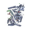
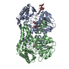
A: UvrABC system protein B
B: UvrABC system protein B
C: UvrABC system protein B
D: DNA (5'-D(*TP*CP*TP*CP*CP*AP*TP*CP*GP*CP*GP*CP*TP*AP*C)-3')
E: DNA (5'-D(*GP*GP*TP*AP*GP*CP*GP*CP*GP*AP*TP*GP*GP*AP*GP*A)-3')
F: DNA (5'-D(*TP*CP*TP*CP*CP*AP*TP*CP*GP*CP*GP*CP*TP*AP*C)-3')
G: DNA (5'-D(*GP*GP*TP*AP*GP*CP*GP*CP*GP*AP*TP*GP*GP*AP*GP*A)-3')
H: DNA (5'-D(*TP*CP*TP*CP*CP*AP*TP*CP*GP*CP*GP*CP*TP*AP*C)-3')
I: DNA (5'-D(*GP*GP*TP*AP*GP*CP*GP*CP*GP*AP*TP*GP*GP*AP*GP*A)-3')
hetero molecules


Theoretical massNumber of molelcules
Total (without water)234,34014
Polymers233,0009
Non-polymers1,3405
Water46826
1
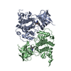
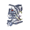
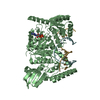
A: UvrABC system protein B
D: DNA (5'-D(*TP*CP*TP*CP*CP*AP*TP*CP*GP*CP*GP*CP*TP*AP*C)-3')
E: DNA (5'-D(*GP*GP*TP*AP*GP*CP*GP*CP*GP*AP*TP*GP*GP*AP*GP*A)-3')
hetero molecules


Theoretical massNumber of molelcules
Total (without water)78,1175
Polymers77,6673
Non-polymers4502
Water181
TypeNameSymmetry operationNumber
identity operation1_555x,y,z1
Buried area5470 Å2
ΔGint-43 kcal/mol
Surface area28610 Å2
MethodPISA
2
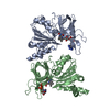
B: UvrABC system protein B
F: DNA (5'-D(*TP*CP*TP*CP*CP*AP*TP*CP*GP*CP*GP*CP*TP*AP*C)-3')
G: DNA (5'-D(*GP*GP*TP*AP*GP*CP*GP*CP*GP*AP*TP*GP*GP*AP*GP*A)-3')
hetero molecules


Theoretical massNumber of molelcules
Total (without water)78,1295
Polymers77,6673
Non-polymers4632
Water181
TypeNameSymmetry operationNumber
identity operation1_555x,y,z1
Buried area5100 Å2
ΔGint-43 kcal/mol
Surface area29320 Å2
MethodPISA
3
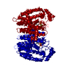
C: UvrABC system protein B
H: DNA (5'-D(*TP*CP*TP*CP*CP*AP*TP*CP*GP*CP*GP*CP*TP*AP*C)-3')
I: DNA (5'-D(*GP*GP*TP*AP*GP*CP*GP*CP*GP*AP*TP*GP*GP*AP*GP*A)-3')
hetero molecules


Theoretical massNumber of molelcules
Total (without water)78,0944
Polymers77,6673
Non-polymers4271
Water181
TypeNameSymmetry operationNumber
identity operation1_555x,y,z1
Buried area5200 Å2
ΔGint-37 kcal/mol
Surface area28960 Å2
MethodPISA
Unit cell
Length a, b, c (Å)170.685, 201.181, 62.664
Angle α, β, γ (deg.)90.000, 90.000, 90.000
Int Tables number18
Space group name H-MP21212

-
Components

-
Protein , 1 types, 3 molecules ABC

#1: Protein UvrABC system protein B / Protein UvrB / Excinuclease ABC subunit B


Mass: 68157.453 Da / Num. of mol.: 3
Source method: isolated from a genetically manipulated source
Source: (gene. exp.) Bacillus caldotenax (bacteria) / Gene: uvrB / Production host: Escherichia coli (E. coli) / References: UniProt: P56981

-
DNA chain , 2 types, 6 molecules DFHEGI

#2: DNA chain DNA (5'-D(*TP*CP*TP*CP*CP*AP*TP*CP*GP*CP*GP*CP*TP*AP*C)-3')


Mass: 4480.911 Da / Num. of mol.: 3 / Source method: obtained synthetically / Source: (synth.) synthetic construct (others)
#3: DNA chain DNA (5'-D(*GP*GP*TP*AP*GP*CP*GP*CP*GP*AP*TP*GP*GP*AP*GP*A)-3')


Mass: 5028.266 Da / Num. of mol.: 3 / Source method: obtained synthetically / Source: (synth.) synthetic construct (others)

-
Non-polymers , 4 types, 31 molecules

#4: Chemical ChemComp-ADP / ADENOSINE-5'-DIPHOSPHATE / Adenosine diphosphate


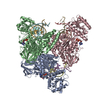
Mass: 427.201 Da / Num. of mol.: 3 / Source method: obtained synthetically / Formula: C10H15N5O10P2 / Comment: ADP, energy-carrying molecule*YM
#5: Chemical ChemComp-CL / CHLORIDE ION / Chloride


Mass: 35.453 Da / Num. of mol.: 1 / Source method: obtained synthetically / Formula: Cl
#6: Chemical ChemComp-NA / SODIUM ION


Mass: 22.990 Da / Num. of mol.: 1 / Source method: obtained synthetically / Formula: Na
#7: Water ChemComp-HOH / water / Water


Mass: 18.015 Da / Num. of mol.: 26 / Source method: isolated from a natural source / Formula: H2O

-
Experimental details

-
Experiment

ExperimentMethod: X-RAY DIFFRACTION / Number of used crystals: 1

-
Sample preparation

CrystalDensity Matthews: 2.31 Å3/Da / Density % sol: 46.8 %
Crystal growTemperature: 293 K / Method: vapor diffusion, sitting drop / pH: 7.5
Details: 0.1 M HEPES, pH 7.5, 0.02 M magnesium chloride, 20-22% poly(acryl acid sodium salt) 5100

-
Data collection

DiffractionMean temperature: 100 K / Serial crystal experiment: N
Diffraction sourceSource: SYNCHROTRON / Site: APS / Beamline: 24-ID-E / Wavelength: 0.9792 Å
DetectorType: ADSC QUANTUM 315 / Detector: CCD / Date: Oct 28, 2011
RadiationMonochromator: Cryogenically-cooled single crystal Si(220) side bounce
Protocol: SINGLE WAVELENGTH / Monochromatic (M) / Laue (L): M / Scattering type: x-ray
Radiation wavelengthWavelength: 0.9792 Å / Relative weight: 1
ReflectionResolution: 2.63→50 Å / Num. obs: 64310 / % possible obs: 99.8 % / Redundancy: 6.2 % / Net I/σ(I): 18.9
Reflection shellResolution: 2.63→2.78 Å / Num. unique obs: 9258

-
Processing

Software
NameVersionClassification
REFMAC5.8.0049refinement
PDB_EXTRACT3.15data extraction
XDSdata reduction
SCALAdata scaling
PHASERphasing
RefinementMethod to determine structure: MOLECULAR REPLACEMENT / Resolution: 2.64→48.24 Å / Cor.coef. Fo:Fc: 0.939 / Cor.coef. Fo:Fc free: 0.906 / SU B: 15.926 / SU ML: 0.326 / Cross valid method: THROUGHOUT / σ(F): 0 / ESU R Free: 0.373 / Stereochemistry target values: MAXIMUM LIKELIHOOD
Details: HYDROGENS HAVE BEEN ADDED IN THE RIDING POSITIONS U VALUES : REFINED INDIVIDUALLY
RfactorNum. reflection% reflectionSelection details
Rfree0.2738 3254 5.1 %RANDOM
Rwork0.22 ---
obs0.2228 61005 99.18 %-
Solvent computationIon probe radii: 0.8 Å / Shrinkage radii: 0.8 Å / VDW probe radii: 1.2 Å / Solvent model: MASK
Displacement parametersBiso max: 155.98 Å2 / Biso mean: 69.098 Å2 / Biso min: 35.96 Å2
Baniso -1Baniso -2Baniso -3
1--0.01 Å20 Å20 Å2
2--0.01 Å20 Å2
3---0 Å2
Refinement stepCycle: final / Resolution: 2.64→48.24 Å
ProteinNucleic acidLigandSolventTotal
Num. atoms13662 1874 83 26 15645
Biso mean--89.32 53.15 -
Num. residues----1784
Refine LS restraints
Refine-IDTypeDev idealDev ideal targetNumber
X-RAY DIFFRACTIONr_bond_refined_d0.0050.01816073
X-RAY DIFFRACTIONr_bond_other_d0.0010.0214658
X-RAY DIFFRACTIONr_angle_refined_deg0.9951.86422115
X-RAY DIFFRACTIONr_angle_other_deg2.364333681
X-RAY DIFFRACTIONr_dihedral_angle_1_deg5.61351685
X-RAY DIFFRACTIONr_dihedral_angle_2_deg33.82823.156713
X-RAY DIFFRACTIONr_dihedral_angle_3_deg15.411152547
X-RAY DIFFRACTIONr_dihedral_angle_4_deg15.24415162
X-RAY DIFFRACTIONr_chiral_restr0.0590.22398
X-RAY DIFFRACTIONr_gen_planes_refined0.0040.0216776
X-RAY DIFFRACTIONr_gen_planes_other0.0020.023708
LS refinement shellResolution: 2.635→2.703 Å / Rfactor Rfree error: 0 / Total num. of bins used: 20
RfactorNum. reflection% reflection
Rfree0.385 226 -
Rwork0.35 4458 -
all-4684 -
obs--99.74 %

+
About Yorodumi

-
News

-
Feb 9, 2022. New format data for meta-information of EMDB entries

New format data for meta-information of EMDB entries

  • Version 3 of the EMDB header file is now the official format.
  • The previous official version 1.9 will be removed from the archive.

Related info.:EMDB header

External links:wwPDB to switch to version 3 of the EMDB data model

-
Aug 12, 2020. Covid-19 info

Covid-19 info

URL: https://pdbj.org/emnavi/covid19.php

New page: Covid-19 featured information page in EM Navigator.

Related info.:Covid-19 info / Mar 5, 2020. Novel coronavirus structure data

+
Mar 5, 2020. Novel coronavirus structure data

Novel coronavirus structure data

Related info.:Yorodumi Speices / Aug 12, 2020. Covid-19 info

External links:COVID-19 featured content - PDBj / Molecule of the Month (242):Coronavirus Proteases

+
Jan 31, 2019. EMDB accession codes are about to change! (news from PDBe EMDB page)

EMDB accession codes are about to change! (news from PDBe EMDB page)

  • The allocation of 4 digits for EMDB accession codes will soon come to an end. Whilst these codes will remain in use, new EMDB accession codes will include an additional digit and will expand incrementally as the available range of codes is exhausted. The current 4-digit format prefixed with “EMD-” (i.e. EMD-XXXX) will advance to a 5-digit format (i.e. EMD-XXXXX), and so on. It is currently estimated that the 4-digit codes will be depleted around Spring 2019, at which point the 5-digit format will come into force.
  • The EM Navigator/Yorodumi systems omit the EMD- prefix.

Related info.:Q: What is EMD? / ID/Accession-code notation in Yorodumi/EM Navigator

External links:EMDB Accession Codes are Changing Soon! / Contact to PDBj

+
Jul 12, 2017. Major update of PDB

Major update of PDB

  • wwPDB released updated PDB data conforming to the new PDBx/mmCIF dictionary.
  • This is a major update changing the version number from 4 to 5, and with Remediation, in which all the entries are updated.
  • In this update, many items about electron microscopy experimental information are reorganized (e.g. em_software).
  • Now, EM Navigator and Yorodumi are based on the updated data.

External links:wwPDB Remediation / Enriched Model Files Conforming to OneDep Data Standards Now Available in the PDB FTP Archive

-
Yorodumi

Thousand views of thousand structures

  • Yorodumi is a browser for structure data from EMDB, PDB, SASBDB, etc.
  • This page is also the successor to EM Navigator detail page, and also detail information page/front-end page for Omokage search.
  • The word "yorodu" (or yorozu) is an old Japanese word meaning "ten thousand". "mi" (miru) is to see.

Related info.:EMDB / PDB / SASBDB / Comparison of 3 databanks / Yorodumi Search / Aug 31, 2016. New EM Navigator & Yorodumi / Yorodumi Papers / Jmol/JSmol / Function and homology information / Changes in new EM Navigator and Yorodumi

Read more