 Movie
Movie Controller

[English] 日本語
 Yorodumi
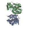
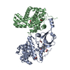
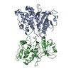
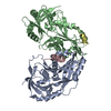
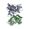
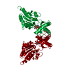
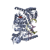
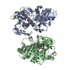
- PDB-1d2m: UVRB PROTEIN OF THERMUS THERMOPHILUS HB8; A NUCLEOTIDE EXCISION R... -
+ Open data

- Basic information
| Entry | Database: PDB / ID: 1d2m | ||||||
|---|---|---|---|---|---|---|---|
| Title | UVRB PROTEIN OF THERMUS THERMOPHILUS HB8; A NUCLEOTIDE EXCISION REPAIR ENZYME | ||||||
 Components Components | EXCINUCLEASE ABC SUBUNIT B | ||||||
 Keywords Keywords | HYDROLASE / MULTIDOMAIN PROTEIN / RIKEN Structural Genomics/Proteomics Initiative / RSGI / Structural Genomics | ||||||
| Function / homology |  Function and homology information Function and homology informationexcinuclease ABC activity / excinuclease repair complex / SOS response / nucleotide-excision repair / ATP hydrolysis activity / DNA binding / ATP binding / cytoplasm Similarity search - Function | ||||||
| Biological species |   Thermus thermophilus (bacteria) Thermus thermophilus (bacteria) | ||||||
| Method |  X-RAY DIFFRACTION / SYNCHROTRON / Resolution: 1.9 Å X-RAY DIFFRACTION / SYNCHROTRON / Resolution: 1.9 Å | ||||||
 Authors Authors | Nakagawa, N. / Sugahara, M. / Masui, R. / Kato, R. / Fukuyama, K. / Kuramitsu, S. / RIKEN Structural Genomics/Proteomics Initiative (RSGI) | ||||||
 Citation Citation | Journal: J.Biochem.(Tokyo) / Year: 1999 Title: Crystal structure of Thermus thermophilus HB8 UvrB protein, a key enzyme of nucleotide excision repair. Authors: Nakagawa, N. / Sugahara, M. / Masui, R. / Kato, R. / Fukuyama, K. / Kuramitsu, S. #1: Journal: Acta Crystallogr.,Sect.D / Year: 1999Title: Crystallization and Preliminary X-ray Diffraction Studies of a DNA Excision Repair Enzyme, UvrB, from Thermus thermophilus HB8 Authors: Shibata, A. / Nakagawa, N. / Sugahara, M. / Masui, R. / Kato, R. / Kuramitsu, S. / Fukuyama, S. | ||||||
| History |
|
- Structure visualization
| Structure viewer | Molecule: MolmilJmol/JSmol |
|---|
- Downloads & links
-Download
| PDBx/mmCIF format | 1d2m.cif.gz | 131.3 KB | Display | PDBx/mmCIF format |
|---|---|---|---|---|
| PDB format | pdb1d2m.ent.gz | 101.8 KB | Display | PDB format |
| PDBx/mmJSON format | 1d2m.json.gz | Tree view | PDBx/mmJSON format | |
| Others |  Other downloads Other downloads |
-Validation report
| Arichive directory | https://data.pdbj.org/pub/pdb/validation_reports/d2/1d2mftp://data.pdbj.org/pub/pdb/validation_reports/d2/1d2m | HTTPS FTP |
|---|
-Related structure data
| Similar structure data | |
|---|---|
| Other databases |


-Links
 PDBj
PDBj- Assembly
| Deposited unit | 
| ||||||||
|---|---|---|---|---|---|---|---|---|---|
| 1 |
| ||||||||
| Unit cell |
|
-Components
| #1: Protein | Mass: 76266.117 Da / Num. of mol.: 1 Source method: isolated from a genetically manipulated source Source: (gene. exp.) Thermus thermophilus (bacteria) / Strain: HB8 / Plasmid: PET11A / Production host: | ||||
|---|---|---|---|---|---|
| #2: Sugar |   Type: D-saccharide / Mass: 292.369 Da / Num. of mol.: 3 / Source method: obtained synthetically / Formula: C14H28O6 / Comment: detergent*YM Type: D-saccharide / Mass: 292.369 Da / Num. of mol.: 3 / Source method: obtained synthetically / Formula: C14H28O6 / Comment: detergent*YM#3: Chemical | ChemComp-SO4 / |   Mass: 96.063 Da / Num. of mol.: 1 / Source method: obtained synthetically / Formula: SO4 Mass: 96.063 Da / Num. of mol.: 1 / Source method: obtained synthetically / Formula: SO4#4: Water | ChemComp-HOH / |  Mass: 18.015 Da / Num. of mol.: 335 / Source method: isolated from a natural source / Formula: H2O Mass: 18.015 Da / Num. of mol.: 335 / Source method: isolated from a natural source / Formula: H2O |
-Experimental details
-Experiment
| Experiment | Method: X-RAY DIFFRACTION / Number of used crystals: 1 |
|---|
- Sample preparation
| Crystal | Density Matthews: 3.68 Å3/Da / Density % sol: 66.6 % | |||||||||||||||||||||||||||||||||||||||||||||
|---|---|---|---|---|---|---|---|---|---|---|---|---|---|---|---|---|---|---|---|---|---|---|---|---|---|---|---|---|---|---|---|---|---|---|---|---|---|---|---|---|---|---|---|---|---|---|
| Crystal grow | Temperature: 293 K / Method: vapor diffusion, hanging drop / pH: 6.1 Details: lithium sulfate, B-octylgulucoside, glycerol, MES, pH 6.1, VAPOR DIFFUSION, HANGING DROP, temperature 293K | |||||||||||||||||||||||||||||||||||||||||||||
| Crystal grow | *PLUS pH: 7.5 / Details: Shibata, A., (1999) Acta Crystallogr.D55, 704. | |||||||||||||||||||||||||||||||||||||||||||||
| Components of the solutions | *PLUS
|
-Data collection
| Diffraction | Mean temperature: 100 K |
|---|---|
| Diffraction source | Source: SYNCHROTRON / Site: SPring-8  / Beamline: BL41XU / Wavelength: 0.708 / Beamline: BL41XU / Wavelength: 0.708 |
| Detector | Type: RIGAKU RAXIS / Detector: IMAGE PLATE / Date: Jun 20, 1998 |
| Radiation | Protocol: SINGLE WAVELENGTH / Monochromatic (M) / Laue (L): M / Scattering type: x-ray |
| Radiation wavelength | Wavelength: 0.708 Å / Relative weight: 1 |
| Reflection | Resolution: 1.9→30 Å / Num. obs: 82751 / % possible obs: 93.5 % / Observed criterion σ(I): 1 / Redundancy: 4 % / Biso Wilson estimate: 15.7 Å2 / Rmerge(I) obs: 0.063 / Net I/σ(I): 21.5 |
| Reflection shell | Resolution: 1.9→1.96 Å / Redundancy: 2.7 % / Rmerge(I) obs: 0.166 / Num. unique all: 3377 / % possible all: 76 |
| Reflection | *PLUS Lowest resolution: 30 Å |
- Processing
| Software |
| ||||||||||||||||||||||||||||||
|---|---|---|---|---|---|---|---|---|---|---|---|---|---|---|---|---|---|---|---|---|---|---|---|---|---|---|---|---|---|---|---|
| Refinement | Resolution: 1.9→30 Å / σ(F): 1 / Stereochemistry target values: ENGH & HUBER
| ||||||||||||||||||||||||||||||
| Refinement step | Cycle: LAST / Resolution: 1.9→30 Å
| ||||||||||||||||||||||||||||||
| Software | *PLUS Name: CNS / Classification: refinement | ||||||||||||||||||||||||||||||
| Refine LS restraints | *PLUS
|
