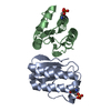
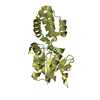
 ムービー
ムービー コントローラー


+ データを開く

- 基本情報
基本情報
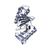
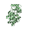
| 登録情報 | データベース: PDB / ID: 3sd7 | ||||||
|---|---|---|---|---|---|---|---|
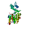
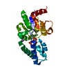
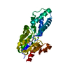
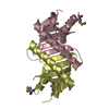
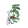
| タイトル | 1.7 Angstrom Resolution Crystal Structure of Putative Phosphatase from Clostridium difficile | ||||||
 要素 要素 | Putative phosphatase | ||||||
 キーワード キーワード | HYDROLASE / Structural Genomics / haloacid dehalogenase-like hydrolase / Center for Structural Genomics of Infectious Diseases / CSGID | ||||||
| 機能・相同性 |  機能・相同性情報 機能・相同性情報加水分解酵素; エステル加水分解酵素; 1価のリン酸エステル加水分解酵素 / protein tyrosine kinase activity / hydrolase activity / cytosol 類似検索 - 分子機能 | ||||||
| 生物種 |  Clostridium difficile (バクテリア) Clostridium difficile (バクテリア) | ||||||
| 手法 |  X線回折 / シンクロトロン / 分子置換 / 解像度: 1.7 Å X線回折 / シンクロトロン / 分子置換 / 解像度: 1.7 Å | ||||||
 データ登録者 データ登録者 | Minasov, G. / Shuvalova, L. / Dubrovska, I. / Winsor, J. / Papazisi, L. / Anderson, W.F. / Center for Structural Genomics of Infectious Diseases (CSGID) | ||||||
 引用 引用 | ジャーナル: Microbiol Resour Announc / 年: 2023 タイトル: A high-throughput structural system biology approach to increase structure representation of proteins from Clostridioides difficile. 著者: Rosas-Lemus, M. / Dey, S. / Minasov, G. / Tan, K. / Anderson, S.M. / Brunzelle, J. / Nocadello, S. / Shabalin, I. / Filippova, E. / Halavaty, A. / Kim, Y. / Maltseva, N. / Osipiuk, J. / ...著者: Rosas-Lemus, M. / Dey, S. / Minasov, G. / Tan, K. / Anderson, S.M. / Brunzelle, J. / Nocadello, S. / Shabalin, I. / Filippova, E. / Halavaty, A. / Kim, Y. / Maltseva, N. / Osipiuk, J. / Minor, W. / Joachimiak, A. / Savchenko, A. / Anderson, W.F. / Satchell, K.J.F. | ||||||
| 履歴 |
|
- 構造の表示
構造の表示
| 構造ビューア | 分子: MolmilJmol/JSmol |
|---|
- ダウンロードとリンク
-ダウンロード
| PDBx/mmCIF形式 | 3sd7.cif.gz | 112 KB | 表示 | PDBx/mmCIF形式 |
|---|---|---|---|---|
| PDB形式 | pdb3sd7.ent.gz | 86.7 KB | 表示 | PDB形式 |
| PDBx/mmJSON形式 | 3sd7.json.gz | ツリー表示 | PDBx/mmJSON形式 | |
| その他 |  その他のダウンロード その他のダウンロード |
-検証レポート
| アーカイブディレクトリ | https://data.pdbj.org/pub/pdb/validation_reports/sd/3sd7ftp://data.pdbj.org/pub/pdb/validation_reports/sd/3sd7 | HTTPS FTP |
|---|
-関連構造データ
| 関連構造データ |  3srtC  3uuwC 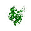 4dd5C  4dgtC  4dq6C  4dunC 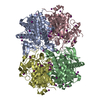 4e1lC 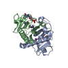 4eguC 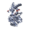 4gibC 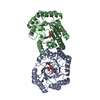 4h3dC  4isxC 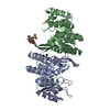 4jjpC 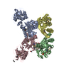 4kd5C  4mfgC  4nmyC  4rn7C 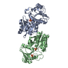 5dzsC  5ttaC 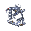 5tv7C 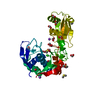 5txuC  6n7mC 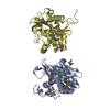 6ue2C  6wy4C  7k1uC  7rl8C  7rlrC 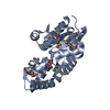 3mc1S S: 精密化の開始モデル C: 同じ文献を引用 ( |
|---|---|
| 類似構造データ | |
| その他のデータベース |
-リンク
 PDBj
PDBj
- 集合体
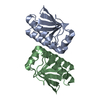
集合体
| 登録構造単位 | 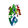
| ||||||||
|---|---|---|---|---|---|---|---|---|---|
| 1 |
| ||||||||
| 単位格子 |
|
-要素
-タンパク質 , 1種, 1分子 A
| #1: タンパク質 | 分子量: 27815.605 Da / 分子数: 1 / 由来タイプ: 組換発現 由来: (組換発現) Clostridium difficile (バクテリア)株: 630 / 遺伝子: CD3605 / プラスミド: pMCSG7 / 発現宿主: |
|---|
-非ポリマー , 5種, 166分子 


| #2: 化合物 | ChemComp-NA /  分子量: 22.990 Da / 分子数: 1 / 由来タイプ: 合成 / 式: Na 分子量: 22.990 Da / 分子数: 1 / 由来タイプ: 合成 / 式: Na | ||
|---|---|---|---|
| #3: 化合物 | ChemComp-CL /  分子量: 35.453 Da / 分子数: 1 / 由来タイプ: 合成 / 式: Cl 分子量: 35.453 Da / 分子数: 1 / 由来タイプ: 合成 / 式: Cl | ||
| #4: 化合物 | ChemComp-PGE /  分子量: 150.173 Da / 分子数: 1 / 由来タイプ: 合成 / 式: C6H14O4 分子量: 150.173 Da / 分子数: 1 / 由来タイプ: 合成 / 式: C6H14O4 | ||
| #5: 化合物 |  分子量: 92.094 Da / 分子数: 2 / 由来タイプ: 合成 / 式: C3H8O3 分子量: 92.094 Da / 分子数: 2 / 由来タイプ: 合成 / 式: C3H8O3#6: 水 | ChemComp-HOH / | 分子量: 18.015 Da / 分子数: 161 / 由来タイプ: 天然 / 式: H2O |
-詳細
| Has protein modification | N |
|---|
-実験情報
-実験
| 実験 | 手法: X線回折 / 使用した結晶の数: 1 |
|---|
- 試料調製
試料調製
| 結晶 | マシュー密度: 2.19 Å3/Da / 溶媒含有率: 43.94 % |
|---|---|
| 結晶化 | 温度: 295 K / 手法: 蒸気拡散法, シッティングドロップ法 / pH: 8.3 詳細: Protein: 7.1mG/mL, 0.5M Sodium chloride, 0.01M Tris, pH 8.3, Screen: JCSG+, D12, 0.04M Potassium Phosphate, 16% (w/v) PEG 8000, 20% (v/v) Glycerol, VAPOR DIFFUSION, SITTING DROP, temperature 295K |
-データ収集
| 放射光源 | 由来: シンクロトロン / サイト: APS  / ビームライン: 21-ID-F / 波長: 0.97872 Å / ビームライン: 21-ID-F / 波長: 0.97872 Å |
|---|---|
| 検出器 | タイプ: MARMOSAIC 225 mm CCD / 検出器: CCD / 日付: 2011年6月6日 / 詳細: Beryllium lenses |
| 放射 | モノクロメーター: Diamond / プロトコル: SINGLE WAVELENGTH / 単色(M)・ラウエ(L): M / 散乱光タイプ: x-ray |
| 放射波長 | 波長: 0.97872 Å / 相対比: 1 |
| 反射 | 解像度: 1.7→30 Å / Num. all: 26648 / Num. obs: 26648 / % possible obs: 99.2 % / Observed criterion σ(I): -3 / 冗長度: 7.2 % / Biso Wilson estimate: 29.7 Å2 / Rmerge(I) obs: 0.048 / Net I/σ(I): 35 |
| 反射 シェル | 解像度: 1.7→1.73 Å / 冗長度: 5.3 % / Rmerge(I) obs: 0.541 / Mean I/σ(I) obs: 2.8 / Num. unique all: 1216 / % possible all: 91.1 |
- 解析
解析
| ソフトウェア |
| |||||||||||||||||||||||||||||||||||||||||||||||||||||||||||||||||||||||||||||||||||||||||||||||||||||||||||||||||||||||||||||
|---|---|---|---|---|---|---|---|---|---|---|---|---|---|---|---|---|---|---|---|---|---|---|---|---|---|---|---|---|---|---|---|---|---|---|---|---|---|---|---|---|---|---|---|---|---|---|---|---|---|---|---|---|---|---|---|---|---|---|---|---|---|---|---|---|---|---|---|---|---|---|---|---|---|---|---|---|---|---|---|---|---|---|---|---|---|---|---|---|---|---|---|---|---|---|---|---|---|---|---|---|---|---|---|---|---|---|---|---|---|---|---|---|---|---|---|---|---|---|---|---|---|---|---|---|---|---|
| 精密化 | 構造決定の手法: 分子置換 開始モデル: 3MC1 解像度: 1.7→29.65 Å / Cor.coef. Fo:Fc: 0.971 / Cor.coef. Fo:Fc free: 0.964 / SU B: 4.094 / SU ML: 0.062 Isotropic thermal model: Thermal Factors Individually Refined 交差検証法: THROUGHOUT / ESU R: 0.1 / ESU R Free: 0.095 / 立体化学のターゲット値: MAXIMUM LIKELIHOOD / 詳細: HYDROGENS HAVE BEEN ADDED IN THE RIDING POSITIONS
| |||||||||||||||||||||||||||||||||||||||||||||||||||||||||||||||||||||||||||||||||||||||||||||||||||||||||||||||||||||||||||||
| 溶媒の処理 | イオンプローブ半径: 0.8 Å / 減衰半径: 0.8 Å / VDWプローブ半径: 1.2 Å / 溶媒モデル: MASK | |||||||||||||||||||||||||||||||||||||||||||||||||||||||||||||||||||||||||||||||||||||||||||||||||||||||||||||||||||||||||||||
| 原子変位パラメータ | Biso mean: 39.35 Å2
| |||||||||||||||||||||||||||||||||||||||||||||||||||||||||||||||||||||||||||||||||||||||||||||||||||||||||||||||||||||||||||||
| 精密化ステップ | サイクル: LAST / 解像度: 1.7→29.65 Å
| |||||||||||||||||||||||||||||||||||||||||||||||||||||||||||||||||||||||||||||||||||||||||||||||||||||||||||||||||||||||||||||
| 拘束条件 |
| |||||||||||||||||||||||||||||||||||||||||||||||||||||||||||||||||||||||||||||||||||||||||||||||||||||||||||||||||||||||||||||
| LS精密化 シェル | 解像度: 1.701→1.745 Å / Total num. of bins used: 20
| |||||||||||||||||||||||||||||||||||||||||||||||||||||||||||||||||||||||||||||||||||||||||||||||||||||||||||||||||||||||||||||
| 精密化 TLS | 手法: refined / Refine-ID: X-RAY DIFFRACTION
| |||||||||||||||||||||||||||||||||||||||||||||||||||||||||||||||||||||||||||||||||||||||||||||||||||||||||||||||||||||||||||||
| 精密化 TLSグループ |
|
