[English] 日本語
Yorodumi
- PDB-6jhk: Crystal Structure of Bacillus subtilis RsbS -

+
Open data


ID or keywords:

Loading...

-
Basic information

Entry
Database: PDB / ID: 6jhk
TitleCrystal Structure of Bacillus subtilis RsbS
ComponentsRsbS negative regulator of sigma-B
KeywordsPROTEIN BINDING / RsbS / stressosome / icosahedron / STAS domain
Function / homology
Function and homology information


STAS domain / Transcription Regulator spoIIAA / STAS domain / STAS domain profile. / STAS domain / STAS domain superfamily / 2-Layer Sandwich / Alpha Beta
Similarity search - Domain/homology
RsbS negative regulator of sigma-B / RsbT antagonist protein RsbS
Similarity search - Component
Biological speciesBacillus subtilis (bacteria)
MethodX-RAY DIFFRACTION / SYNCHROTRON / SAD / Resolution: 3.101 Å
Model detailsCrystal structure of icosahedral BsRsbS
AuthorsKwon, E. / Pathak, D. / Dahal, P. / Kim, D.Y.
Funding support Korea, Republic Of, 1items
OrganizationGrant numberCountry
National Research Foundation (Korea) Korea, Republic Of
Citation
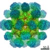
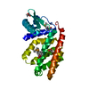
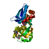
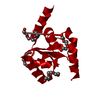
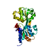
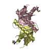
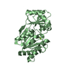
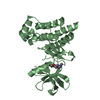
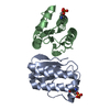
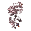
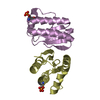
Journal: IUCrJ / Year: 2019
Title: Structural insights into stressosome assembly.
Authors: Eunju Kwon / Deepak Pathak / Han-Ul Kim / Pawan Dahal / Sung Chul Ha / Seung Sik Lee / Hyeongseop Jeong / Dooil Jeoung / Hyeun Wook Chang / Hyun Suk Jung / Dong Young Kim /
Abstract: The stressosome transduces environmental stress signals to SigB to upregulate SigB-dependent transcription, which is required for bacterial viability. The stressosome core is composed of RsbS and at ...The stressosome transduces environmental stress signals to SigB to upregulate SigB-dependent transcription, which is required for bacterial viability. The stressosome core is composed of RsbS and at least one of the RsbR paralogs. A previous cryo-electron microscopy (cryo-EM) structure of the RsbRA-RsbS complex determined under a 2 symmetry restraint showed that the stressosome core forms a pseudo-icosahedron consisting of 60 STAS domains of RsbRA and RsbS. However, it is still unclear how RsbS and one of the RsbR paralogs assemble into the stressosome. Here, an assembly model of the stressosome is presented based on the crystal structure of the RsbS icosahedron and cryo-EM structures of the RsbRA-RsbS complex determined under diverse symmetry restraints (nonsymmetric 1, dihedral 2 and icosahedral envelopes). 60 monomers of the crystal structure of RsbS fitted well into the -restrained cryo-EM structure determined at 4.1 Å resolution, even though the STAS domains in the envelope were averaged. This indicates that RsbS and RsbRA share a highly conserved STAS fold. 22 protrusions observed in the 1 envelope, corresponding to dimers of the RsbRA N-domain, allowed the STAS domains of RsbRA and RsbS to be distinguished in the stressosome core. Based on these, the model of the stressosome core was reconstructed. The mutation of RsbRA residues at the binding interface in the model (R189A/Q191A) significantly reduced the interaction between RsbRA and RsbS. These results suggest that nonconserved residues in the conserved STAS folds between RsbS and RsbR paralogs determine stressosome assembly.
History
DepositionFeb 18, 2019Deposition site: PDBJ / Processing site: PDBJ
Revision 1.0Sep 11, 2019Provider: repository / Type: Initial release
Revision 1.1Nov 20, 2019Group: Database references / Category: citation / citation_author
Item: _citation.pdbx_database_id_PubMed / _citation.title / _citation_author.name

-
Structure visualization

Structure viewerMolecule:
MolmilJmol/JSmol

Downloads & links

-
Assembly

Deposited unit
A: RsbS negative regulator of sigma-B
B: RsbS negative regulator of sigma-B
C: RsbS negative regulator of sigma-B
D: RsbS negative regulator of sigma-B
E: RsbS negative regulator of sigma-B


Theoretical massNumber of molelcules
Total (without water)67,7235
Polymers67,7235
Non-polymers00
Water00
1
A: RsbS negative regulator of sigma-B

B: RsbS negative regulator of sigma-B


Theoretical massNumber of molelcules
Total (without water)27,0892
Polymers27,0892
Non-polymers00
Water0
TypeNameSymmetry operationNumber
identity operation1_555x,y,z1
crystal symmetry operation12_665-y+1,-z+1,x1
Buried area1450 Å2
ΔGint-8 kcal/mol
Surface area11680 Å2
2
C: RsbS negative regulator of sigma-B

C: RsbS negative regulator of sigma-B


Theoretical massNumber of molelcules
Total (without water)27,0892
Polymers27,0892
Non-polymers00
Water0
TypeNameSymmetry operationNumber
identity operation1_555x,y,z1
crystal symmetry operation2_755-x+2,-y,z1
Buried area1500 Å2
ΔGint-8 kcal/mol
Surface area11630 Å2
3
D: RsbS negative regulator of sigma-B

E: RsbS negative regulator of sigma-B


Theoretical massNumber of molelcules
Total (without water)27,0892
Polymers27,0892
Non-polymers00
Water0
TypeNameSymmetry operationNumber
identity operation1_555x,y,z1
crystal symmetry operation8_746-z+2,x-1,-y+11
Buried area1490 Å2
ΔGint-10 kcal/mol
Surface area11500 Å2
4


  • Idetical with deposited unit
  • defined by software
TypeNameSymmetry operationNumber
identity operation1_555x,y,z1
Buried area6340 Å2
ΔGint-58 kcal/mol
Surface area26350 Å2
MethodPISA
Unit cell
Length a, b, c (Å)179.201, 179.201, 179.201
Angle α, β, γ (deg.)90.000, 90.000, 90.000
Int Tables number197
Space group name H-MI23

-
Components

#1: Protein
RsbS negative regulator of sigma-B / RsbT antagonist protein RsbS


Mass: 13544.638 Da / Num. of mol.: 5
Source method: isolated from a genetically manipulated source
Source: (gene. exp.) Bacillus subtilis (bacteria)
Gene: B4122_0858, B4417_4413, CJ481_02205, ETA10_02695, ETK61_02665, ETK71_02600
Plasmid: pETDuet-1 / Production host: Escherichia coli (E. coli) / Strain (production host): BL21 Star(DE3) / References: UniProt: A0A063XHI7, UniProt: P42410*PLUS

-
Experimental details

-
Experiment

ExperimentMethod: X-RAY DIFFRACTION / Number of used crystals: 1

-
Sample preparation

CrystalDensity Matthews: 3.75 Å3/Da / Density % sol: 67.2 %
Crystal growTemperature: 295 K / Method: batch mode / pH: 6 / Details: MPD, LiSO4

-
Data collection

DiffractionMean temperature: 100 K / Serial crystal experiment: N
Diffraction sourceSource: SYNCHROTRON / Site: PAL/PLS / Beamline: 7A (6B, 6C1) / Wavelength: 0.97933 Å
DetectorType: ADSC QUANTUM 270 / Detector: CCD / Date: Dec 15, 2016
RadiationProtocol: SINGLE WAVELENGTH / Monochromatic (M) / Laue (L): M / Scattering type: x-ray
Radiation wavelengthWavelength: 0.97933 Å / Relative weight: 1
ReflectionResolution: 3.1→30 Å / Num. obs: 17310 / % possible obs: 98.9 % / Redundancy: 5.97 % / Biso Wilson estimate: 92.68 Å2 / Rmerge(I) obs: 0.072 / Net I/σ(I): 38.9
Reflection shellResolution: 3.1→3.15 Å / Rmerge(I) obs: 0.546 / Mean I/σ(I) obs: 5.2 / Num. unique obs: 851 / % possible all: 100

-
Processing

Software
NameVersionClassification
HKL-2000data reduction
SCALEPACKdata scaling
PHENIXdev_2313refinement
PDB_EXTRACT3.24data extraction
PHENIXphasing
RefinementMethod to determine structure: SAD / Resolution: 3.101→29.867 Å / SU ML: 0.34 / Cross valid method: THROUGHOUT / σ(F): 1.34 / Phase error: 24.37
RfactorNum. reflection% reflection
Rfree0.2405 1732 10.02 %
Rwork0.2102 --
obs0.2131 17280 98.8 %
Solvent computationShrinkage radii: 0.9 Å / VDW probe radii: 1.11 Å
Displacement parametersBiso max: 145.96 Å2 / Biso mean: 82.3339 Å2 / Biso min: 51.25 Å2
Refinement stepCycle: final / Resolution: 3.101→29.867 Å
ProteinNucleic acidLigandSolventTotal
Num. atoms4437 0 0 0 4437
Num. residues----576
Refine LS restraints
Refine-IDTypeDev idealNumber
X-RAY DIFFRACTIONf_bond_d0.0044473
X-RAY DIFFRACTIONf_angle_d0.7176081
X-RAY DIFFRACTIONf_chiral_restr0.049796
X-RAY DIFFRACTIONf_plane_restr0.003741
X-RAY DIFFRACTIONf_dihedral_angle_d13.2772741
LS refinement shell

Refine-ID: X-RAY DIFFRACTION / Rfactor Rfree error: 0 / Total num. of bins used: 12

Resolution (Å)Rfactor RfreeNum. reflection RfreeRfactor RworkNum. reflection RworkNum. reflection all% reflection obs (%)
3.1008-3.19190.32181450.287912851430100
3.1919-3.29480.35321400.293912991439100
3.2948-3.41240.29351450.284312891434100
3.4124-3.54880.31581470.260513171464100
3.5488-3.710.22961480.217712991447100
3.71-3.90520.24091420.225213141456100
3.9052-4.14930.24871490.2212871436100
4.1493-4.46870.21321440.18213011445100
4.4687-4.91660.20281490.18513091458100
4.9166-5.6240.23471510.204613211472100
5.624-7.07010.2551450.225613301475100
7.0701-29.86820.20711270.16971197132487

+
About Yorodumi

-
News

-
Feb 9, 2022. New format data for meta-information of EMDB entries

New format data for meta-information of EMDB entries

  • Version 3 of the EMDB header file is now the official format.
  • The previous official version 1.9 will be removed from the archive.

Related info.:EMDB header

External links:wwPDB to switch to version 3 of the EMDB data model

-
Aug 12, 2020. Covid-19 info

Covid-19 info

URL: https://pdbjlvh1.pdbj.org/emnavi/covid19.php

New page: Covid-19 featured information page in EM Navigator.

Related info.:Covid-19 info / Mar 5, 2020. Novel coronavirus structure data

+
Mar 5, 2020. Novel coronavirus structure data

Novel coronavirus structure data

Related info.:Yorodumi Speices / Aug 12, 2020. Covid-19 info

External links:COVID-19 featured content - PDBj / Molecule of the Month (242):Coronavirus Proteases

+
Jan 31, 2019. EMDB accession codes are about to change! (news from PDBe EMDB page)

EMDB accession codes are about to change! (news from PDBe EMDB page)

  • The allocation of 4 digits for EMDB accession codes will soon come to an end. Whilst these codes will remain in use, new EMDB accession codes will include an additional digit and will expand incrementally as the available range of codes is exhausted. The current 4-digit format prefixed with “EMD-” (i.e. EMD-XXXX) will advance to a 5-digit format (i.e. EMD-XXXXX), and so on. It is currently estimated that the 4-digit codes will be depleted around Spring 2019, at which point the 5-digit format will come into force.
  • The EM Navigator/Yorodumi systems omit the EMD- prefix.

Related info.:Q: What is EMD? / ID/Accession-code notation in Yorodumi/EM Navigator

External links:EMDB Accession Codes are Changing Soon! / Contact to PDBj

+
Jul 12, 2017. Major update of PDB

Major update of PDB

  • wwPDB released updated PDB data conforming to the new PDBx/mmCIF dictionary.
  • This is a major update changing the version number from 4 to 5, and with Remediation, in which all the entries are updated.
  • In this update, many items about electron microscopy experimental information are reorganized (e.g. em_software).
  • Now, EM Navigator and Yorodumi are based on the updated data.

External links:wwPDB Remediation / Enriched Model Files Conforming to OneDep Data Standards Now Available in the PDB FTP Archive

-
Yorodumi

Thousand views of thousand structures

  • Yorodumi is a browser for structure data from EMDB, PDB, SASBDB, etc.
  • This page is also the successor to EM Navigator detail page, and also detail information page/front-end page for Omokage search.
  • The word "yorodu" (or yorozu) is an old Japanese word meaning "ten thousand". "mi" (miru) is to see.

Related info.:EMDB / PDB / SASBDB / Comparison of 3 databanks / Yorodumi Search / Aug 31, 2016. New EM Navigator & Yorodumi / Yorodumi Papers / Jmol/JSmol / Function and homology information / Changes in new EM Navigator and Yorodumi

Read more