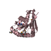
 Movie
Movie Controller

[English] 日本語
 Yorodumi
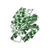
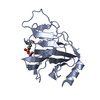
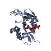
- PDB-4rn7: The crystal structure of N-acetylmuramoyl-L-alanine amidase from ... -
+ Open data

- Basic information
| Entry | Database: PDB / ID: 4rn7 | ||||||
|---|---|---|---|---|---|---|---|
| Title | The crystal structure of N-acetylmuramoyl-L-alanine amidase from Clostridium difficile 630 | ||||||
 Components Components | N-acetylmuramoyl-L-alanine amidase | ||||||
 Keywords Keywords | HYDROLASE / structural genomics / Center for Structural Genomics of Infectious Diseases / CSGID | ||||||
| Function / homology |  Function and homology information Function and homology informationN-acetylmuramoyl-L-alanine amidase / N-acetylmuramoyl-L-alanine amidase activity / peptidoglycan catabolic process / outer membrane-bounded periplasmic space / membrane / metal ion binding Similarity search - Function | ||||||
| Biological species |  Peptoclostridium difficile (bacteria) Peptoclostridium difficile (bacteria) | ||||||
| Method |  X-RAY DIFFRACTION / SYNCHROTRON / SAD / Resolution: 1.717 Å X-RAY DIFFRACTION / SYNCHROTRON / SAD / Resolution: 1.717 Å | ||||||
 Authors Authors | Tan, K. / Mulligan, R. / Kwon, K. / Anderson, W.F. / Joachimiak, A. / Center for Structural Genomics of Infectious Diseases (CSGID) | ||||||
 Citation Citation | Journal: Microbiol Resour Announc / Year: 2023 Title: A high-throughput structural system biology approach to increase structure representation of proteins from Clostridioides difficile. Authors: Rosas-Lemus, M. / Dey, S. / Minasov, G. / Tan, K. / Anderson, S.M. / Brunzelle, J. / Nocadello, S. / Shabalin, I. / Filippova, E. / Halavaty, A. / Kim, Y. / Maltseva, N. / Osipiuk, J. / ...Authors: Rosas-Lemus, M. / Dey, S. / Minasov, G. / Tan, K. / Anderson, S.M. / Brunzelle, J. / Nocadello, S. / Shabalin, I. / Filippova, E. / Halavaty, A. / Kim, Y. / Maltseva, N. / Osipiuk, J. / Minor, W. / Joachimiak, A. / Savchenko, A. / Anderson, W.F. / Satchell, K.J.F. | ||||||
| History |
|
- Structure visualization
| Structure viewer | Molecule: MolmilJmol/JSmol |
|---|
- Downloads & links
-Download
| PDBx/mmCIF format | 4rn7.cif.gz | 93.8 KB | Display | PDBx/mmCIF format |
|---|---|---|---|---|
| PDB format | pdb4rn7.ent.gz | 70.4 KB | Display | PDB format |
| PDBx/mmJSON format | 4rn7.json.gz | Tree view | PDBx/mmJSON format | |
| Others |  Other downloads Other downloads |
-Validation report
| Arichive directory | https://data.pdbj.org/pub/pdb/validation_reports/rn/4rn7ftp://data.pdbj.org/pub/pdb/validation_reports/rn/4rn7 | HTTPS FTP |
|---|
-Related structure data
| Related structure data | 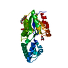 3sd7C  3srtC  3uuwC 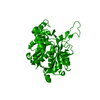 4dd5C  4dgtC  4dq6C  4dunC 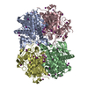 4e1lC 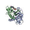 4eguC 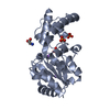 4gibC 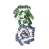 4h3dC  4isxC 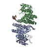 4jjpC 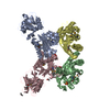 4kd5C  4mfgC  4nmyC 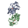 5dzsC  5ttaC 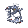 5tv7C 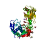 5txuC 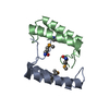 6n7mC 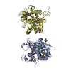 6ue2C  6wy4C  7k1uC  7rl8C  7rlrC C: citing same article ( |
|---|---|
| Similar structure data | |
| Other databases |


-Links
 PDBj
PDBj- Assembly
| Deposited unit | 
| ||||||||
|---|---|---|---|---|---|---|---|---|---|
| 1 |
| ||||||||
| Unit cell |
|
-Components
-Protein , 1 types, 1 molecules A
| #1: Protein | Mass: 20886.156 Da / Num. of mol.: 1 / Fragment: UNP residues 117-301 Source method: isolated from a genetically manipulated source Source: (gene. exp.) Peptoclostridium difficile (bacteria) / Strain: 630 / Gene: CD630_27610 / Plasmid: pMCSG7 / Production host: References: UniProt: Q183J9, N-acetylmuramoyl-L-alanine amidase |
|---|
-Non-polymers , 5 types, 129 molecules 


| #2: Chemical | ChemComp-ZN /  Mass: 65.409 Da / Num. of mol.: 1 / Source method: obtained synthetically / Formula: Zn Mass: 65.409 Da / Num. of mol.: 1 / Source method: obtained synthetically / Formula: Zn | ||||
|---|---|---|---|---|---|
| #3: Chemical | ChemComp-GOL /  Mass: 92.094 Da / Num. of mol.: 1 / Source method: obtained synthetically / Formula: C3H8O3 Mass: 92.094 Da / Num. of mol.: 1 / Source method: obtained synthetically / Formula: C3H8O3 | ||||
| #4: Chemical |  Mass: 238.305 Da / Num. of mol.: 2 / Source method: obtained synthetically / Formula: C8H18N2O4S / Comment: pH buffer*YM Mass: 238.305 Da / Num. of mol.: 2 / Source method: obtained synthetically / Formula: C8H18N2O4S / Comment: pH buffer*YM#5: Chemical |  Mass: 46.025 Da / Num. of mol.: 2 / Source method: obtained synthetically / Formula: CH2O2 Mass: 46.025 Da / Num. of mol.: 2 / Source method: obtained synthetically / Formula: CH2O2#6: Water | ChemComp-HOH / | Mass: 18.015 Da / Num. of mol.: 123 / Source method: isolated from a natural source / Formula: H2O |
-Details
| Has protein modification | Y |
|---|
-Experimental details
-Experiment
| Experiment | Method: X-RAY DIFFRACTION / Number of used crystals: 1 |
|---|
- Sample preparation
| Crystal | Density Matthews: 2.93 Å3/Da / Density % sol: 58.04 % |
|---|---|
| Crystal grow | Temperature: 289 K / Method: vapor diffusion, sitting drop / pH: 7.5 Details: 0.1M HEPES, 25% w/v PEG3350, pH 7.5, VAPOR DIFFUSION, SITTING DROP, temperature 289K |
-Data collection
| Diffraction | Mean temperature: 100 K |
|---|---|
| Diffraction source | Source: SYNCHROTRON / Site: APS  / Beamline: 19-ID / Wavelength: 0.97899 Å / Beamline: 19-ID / Wavelength: 0.97899 Å |
| Detector | Type: ADSC QUANTUM 315r / Detector: CCD / Date: Mar 18, 2013 / Details: mirror |
| Radiation | Monochromator: Si 111 crystal / Protocol: SINGLE WAVELENGTH / Monochromatic (M) / Laue (L): M / Scattering type: x-ray |
| Radiation wavelength | Wavelength: 0.97899 Å / Relative weight: 1 |
| Reflection | Resolution: 1.72→28.8 Å / Num. all: 26769 / Num. obs: 26769 / % possible obs: 99.8 % / Observed criterion σ(F): 0 / Observed criterion σ(I): -5 / Redundancy: 8.3 % / Biso Wilson estimate: 27.45 Å2 / Rmerge(I) obs: 0.055 / Net I/σ(I): 54.7 |
| Reflection shell | Resolution: 1.72→1.75 Å / Redundancy: 8.4 % / Rmerge(I) obs: 0.717 / Mean I/σ(I) obs: 3.3 / Num. unique all: 1344 / % possible all: 100 |
- Processing
| Software |
| |||||||||||||||||||||||||||||||||||||||||||||||||||||||||||||||||||||||||||||||||||||||||||||||||||||||||||||||||||||||||||||
|---|---|---|---|---|---|---|---|---|---|---|---|---|---|---|---|---|---|---|---|---|---|---|---|---|---|---|---|---|---|---|---|---|---|---|---|---|---|---|---|---|---|---|---|---|---|---|---|---|---|---|---|---|---|---|---|---|---|---|---|---|---|---|---|---|---|---|---|---|---|---|---|---|---|---|---|---|---|---|---|---|---|---|---|---|---|---|---|---|---|---|---|---|---|---|---|---|---|---|---|---|---|---|---|---|---|---|---|---|---|---|---|---|---|---|---|---|---|---|---|---|---|---|---|---|---|---|
| Refinement | Method to determine structure: SAD / Resolution: 1.717→28.755 Å / SU ML: 0.17 / σ(F): 1.34 / Phase error: 23.97 / Stereochemistry target values: ML
| |||||||||||||||||||||||||||||||||||||||||||||||||||||||||||||||||||||||||||||||||||||||||||||||||||||||||||||||||||||||||||||
| Solvent computation | Shrinkage radii: 0.9 Å / VDW probe radii: 1.11 Å / Solvent model: FLAT BULK SOLVENT MODEL | |||||||||||||||||||||||||||||||||||||||||||||||||||||||||||||||||||||||||||||||||||||||||||||||||||||||||||||||||||||||||||||
| Refinement step | Cycle: LAST / Resolution: 1.717→28.755 Å
| |||||||||||||||||||||||||||||||||||||||||||||||||||||||||||||||||||||||||||||||||||||||||||||||||||||||||||||||||||||||||||||
| Refine LS restraints |
| |||||||||||||||||||||||||||||||||||||||||||||||||||||||||||||||||||||||||||||||||||||||||||||||||||||||||||||||||||||||||||||
| LS refinement shell |
| |||||||||||||||||||||||||||||||||||||||||||||||||||||||||||||||||||||||||||||||||||||||||||||||||||||||||||||||||||||||||||||
| Refinement TLS params. | Method: refined / Refine-ID: X-RAY DIFFRACTION
| |||||||||||||||||||||||||||||||||||||||||||||||||||||||||||||||||||||||||||||||||||||||||||||||||||||||||||||||||||||||||||||
| Refinement TLS group |
|
