 Movie
Movie Controller

[English] 日本語
 Yorodumi
- PDB-6r73: Structure of IMP-13 metallo-beta-lactamase complexed with hydroly... -
+ Open data

- Basic information
| Entry | Database: PDB / ID: 6r73 | ||||||||||||
|---|---|---|---|---|---|---|---|---|---|---|---|---|---|
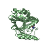
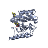
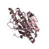
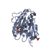
| Title | Structure of IMP-13 metallo-beta-lactamase complexed with hydrolysed meropenem | ||||||||||||
 Components Components | Beta-lactamase | ||||||||||||
 Keywords Keywords | HYDROLASE / Metallo-Beta-Lactamase / Zinc-binding protein / antibiotic resistance | ||||||||||||
| Function / homology |  Function and homology information Function and homology informationantibiotic catabolic process / beta-lactamase activity / beta-lactamase / periplasmic space / response to antibiotic / zinc ion binding Similarity search - Function | ||||||||||||
| Biological species |   Pseudomonas aeruginosa (bacteria) Pseudomonas aeruginosa (bacteria) | ||||||||||||
| Method |  X-RAY DIFFRACTION / SYNCHROTRON / MOLECULAR REPLACEMENT / Resolution: 2.3 Å X-RAY DIFFRACTION / SYNCHROTRON / MOLECULAR REPLACEMENT / Resolution: 2.3 Å | ||||||||||||
 Authors Authors | Softley, C.A. / Zak, K. / Kolonko, M. / Sattler, M. / Popowicz, G. | ||||||||||||
| Funding support |  Germany, Germany,  Poland, 3items Poland, 3items
| ||||||||||||
 Citation Citation | Journal: Antimicrob.Agents Chemother. / Year: 2020 Title: Structure and Molecular Recognition Mechanism of IMP-13 Metallo-beta-Lactamase. Authors: Softley, C.A. / Zak, K.M. / Bostock, M.J. / Fino, R. / Zhou, R.X. / Kolonko, M. / Mejdi-Nitiu, R. / Meyer, H. / Sattler, M. / Popowicz, G.M. | ||||||||||||
| History |
|
- Structure visualization
| Structure viewer | Molecule: MolmilJmol/JSmol |
|---|
- Downloads & links
-Download
| PDBx/mmCIF format | 6r73.cif.gz | 101.1 KB | Display | PDBx/mmCIF format |
|---|---|---|---|---|
| PDB format | pdb6r73.ent.gz | 75 KB | Display | PDB format |
| PDBx/mmJSON format | 6r73.json.gz | Tree view | PDBx/mmJSON format | |
| Others |  Other downloads Other downloads |
-Validation report
| Arichive directory | https://data.pdbj.org/pub/pdb/validation_reports/r7/6r73ftp://data.pdbj.org/pub/pdb/validation_reports/r7/6r73 | HTTPS FTP |
|---|
-Related structure data
| Related structure data |  6r78C  6r79C 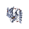 6rzrC  6rzsC  6s0hC  1dd6S S: Starting model for refinement C: citing same article ( |
|---|---|
| Similar structure data |


-Links
 PDBj
PDBj

- Assembly
| Deposited unit | 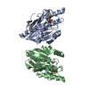
| ||||||||
|---|---|---|---|---|---|---|---|---|---|
| 1 | 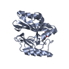
| ||||||||
| 2 | 
| ||||||||
| Unit cell |
|
-Components
| #1: Protein | Mass: 25110.533 Da / Num. of mol.: 2 Source method: isolated from a genetically manipulated source Source: (gene. exp.) Pseudomonas aeruginosa (bacteria) / Gene: bla-imp13, bla-IMP13, blaIMP-13 / Production host: #2: Chemical |   Mass: 401.478 Da / Num. of mol.: 2 / Source method: obtained synthetically / Formula: C17H27N3O6S / Feature type: SUBJECT OF INVESTIGATION Mass: 401.478 Da / Num. of mol.: 2 / Source method: obtained synthetically / Formula: C17H27N3O6S / Feature type: SUBJECT OF INVESTIGATION#3: Chemical | ChemComp-ZN /   Mass: 65.409 Da / Num. of mol.: 4 / Source method: obtained synthetically / Formula: Zn Mass: 65.409 Da / Num. of mol.: 4 / Source method: obtained synthetically / Formula: Zn#4: Water | ChemComp-HOH / |  Mass: 18.015 Da / Num. of mol.: 98 / Source method: isolated from a natural source / Formula: H2O Mass: 18.015 Da / Num. of mol.: 98 / Source method: isolated from a natural source / Formula: H2OHas protein modification | Y | |
|---|
-Experimental details
-Experiment
| Experiment | Method: X-RAY DIFFRACTION / Number of used crystals: 1 |
|---|
- Sample preparation
| Crystal | Density Matthews: 2.42 Å3/Da / Density % sol: 49.27 % |
|---|---|
| Crystal grow | Temperature: 298 K / Method: vapor diffusion, sitting drop / pH: 6.5 Details: 0.1 M Bis-Tris pH 6.5, 25% PEG 3350, cryo-protected with MPD |
-Data collection
| Diffraction | Mean temperature: 90 K / Serial crystal experiment: N |
|---|---|
| Diffraction source | Source: SYNCHROTRON / Site: ESRF  / Beamline: ID30B / Wavelength: 0.9765 Å / Beamline: ID30B / Wavelength: 0.9765 Å |
| Detector | Type: DECTRIS PILATUS3 S 6M / Detector: PIXEL / Date: Jun 9, 2018 |
| Radiation | Monochromator: Si(111) / Protocol: SINGLE WAVELENGTH / Monochromatic (M) / Laue (L): M / Scattering type: x-ray |
| Radiation wavelength | Wavelength: 0.9765 Å / Relative weight: 1 |
| Reflection | Resolution: 2.3→58.04 Å / Num. obs: 20856 / % possible obs: 97.01 % / Redundancy: 1.9 % / Biso Wilson estimate: 33.2 Å2 / CC1/2: 0.995 / Rmerge(I) obs: 0.06752 / Net I/σ(I): 7.62 |
| Reflection shell | Resolution: 2.3→2.382 Å / Redundancy: 1.9 % / Rmerge(I) obs: 0.402 / Mean I/σ(I) obs: 1.94 / Num. unique obs: 2130 / CC1/2: 0.84 / % possible all: 99.3 |
- Processing
| Software |
| |||||||||||||||||||||||||||||||||||||||||||||||||||||||||||||||
|---|---|---|---|---|---|---|---|---|---|---|---|---|---|---|---|---|---|---|---|---|---|---|---|---|---|---|---|---|---|---|---|---|---|---|---|---|---|---|---|---|---|---|---|---|---|---|---|---|---|---|---|---|---|---|---|---|---|---|---|---|---|---|---|---|
| Refinement | Method to determine structure: MOLECULAR REPLACEMENT Starting model: 1DD6 Resolution: 2.3→58.04 Å / SU ML: 0.33 / Cross valid method: FREE R-VALUE / σ(F): 1.35 / Phase error: 30.51
| |||||||||||||||||||||||||||||||||||||||||||||||||||||||||||||||
| Solvent computation | Shrinkage radii: 0.9 Å / VDW probe radii: 1.11 Å | |||||||||||||||||||||||||||||||||||||||||||||||||||||||||||||||
| Refinement step | Cycle: LAST / Resolution: 2.3→58.04 Å
| |||||||||||||||||||||||||||||||||||||||||||||||||||||||||||||||
| Refine LS restraints |
| |||||||||||||||||||||||||||||||||||||||||||||||||||||||||||||||
| LS refinement shell |
|
