 Movie
Movie Controller

[English] 日本語
 Yorodumi
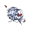
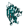
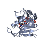
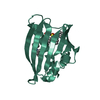
- PDB-7cw1: Crystal structure of a biodegradable plastic-degrading cutinase-l... -
+ Open data

- Basic information
| Entry | Database: PDB / ID: 7cw1 | ||||||
|---|---|---|---|---|---|---|---|
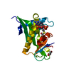
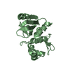
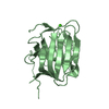
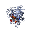
| Title | Crystal structure of a biodegradable plastic-degrading cutinase-like enzyme from the phyllosphere yeast, Pseudozyma antarctica, solved by getting the phase from anomalous scattering of uncovalently coordinated arsenic (cacodylate). | ||||||
 Components Components | Cutinase-like enzyme | ||||||
 Keywords Keywords | HYDROLASE / cutinase-like enzyme / biodegradable plastic degrading enzyme / alpha/beta hydrolase fold | ||||||
| Function / homology | Cutinase / Cutinase/acetylxylan esterase / Cutinase / carboxylic ester hydrolase activity / Alpha/Beta hydrolase fold / CACODYLIC ACID / Cutinase-like enzyme Function and homology information Function and homology information | ||||||
| Biological species |  Pseudozyma antarctica (fungus) Pseudozyma antarctica (fungus) | ||||||
| Method |  X-RAY DIFFRACTION / SYNCHROTRON / SAD / Resolution: 1.7 Å X-RAY DIFFRACTION / SYNCHROTRON / SAD / Resolution: 1.7 Å | ||||||
 Authors Authors | Suzuki, K. | ||||||
 Citation Citation | Journal: To Be Published Title: Crystal structure of a biodegradable plastic-degrading cutinase-like enzyme from the phyllosphere yeast, Pseudozyma antarctica, solved by getting the phase from anomalous scattering of ...Title: Crystal structure of a biodegradable plastic-degrading cutinase-like enzyme from the phyllosphere yeast, Pseudozyma antarctica, solved by getting the phase from anomalous scattering of uncovalently coordinated arsenic (cacodylate). Authors: Suzuki, K. | ||||||
| History |
|
- Structure visualization
| Structure viewer | Molecule: MolmilJmol/JSmol |
|---|
- Downloads & links
-Download
| PDBx/mmCIF format | 7cw1.cif.gz | 91.3 KB | Display | PDBx/mmCIF format |
|---|---|---|---|---|
| PDB format | pdb7cw1.ent.gz | 68.3 KB | Display | PDB format |
| PDBx/mmJSON format | 7cw1.json.gz | Tree view | PDBx/mmJSON format | |
| Others |  Other downloads Other downloads |
-Validation report
| Arichive directory | https://data.pdbj.org/pub/pdb/validation_reports/cw/7cw1ftp://data.pdbj.org/pub/pdb/validation_reports/cw/7cw1 | HTTPS FTP |
|---|
-Related structure data
| Related structure data | |
|---|---|
| Similar structure data |


-Links
 PDBj
PDBj- Assembly
| Deposited unit | 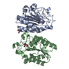
| ||||||||
|---|---|---|---|---|---|---|---|---|---|
| 1 | 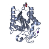
| ||||||||
| 2 | 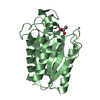
| ||||||||
| Unit cell |
|
-Components
| #1: Protein | Mass: 20376.658 Da / Num. of mol.: 2 / Source method: isolated from a natural source / Source: (natural) Pseudozyma antarctica (fungus) / References: UniProt: S6BC01#2: Chemical | 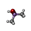  Mass: 137.997 Da / Num. of mol.: 2 / Source method: obtained synthetically / Formula: C2H7AsO2 / Feature type: SUBJECT OF INVESTIGATION Mass: 137.997 Da / Num. of mol.: 2 / Source method: obtained synthetically / Formula: C2H7AsO2 / Feature type: SUBJECT OF INVESTIGATION#3: Chemical | ChemComp-NA / |   Mass: 22.990 Da / Num. of mol.: 1 / Source method: obtained synthetically / Formula: Na Mass: 22.990 Da / Num. of mol.: 1 / Source method: obtained synthetically / Formula: Na#4: Water | ChemComp-HOH / |  Mass: 18.015 Da / Num. of mol.: 361 / Source method: isolated from a natural source / Formula: H2O Mass: 18.015 Da / Num. of mol.: 361 / Source method: isolated from a natural source / Formula: H2OHas ligand of interest | Y | Has protein modification | Y | |
|---|
-Experimental details
-Experiment
| Experiment | Method: X-RAY DIFFRACTION / Number of used crystals: 1 |
|---|
- Sample preparation
| Crystal | Density Matthews: 1.839 Å3/Da / Density % sol: 33.157 % |
|---|---|
| Crystal grow | Temperature: 293 K / Method: vapor diffusion, hanging drop / pH: 6.5 Details: 0.17M Sodium acetate trihydrate, 0.085M Sodium cacodylate trihydrate , 25.5%w/v Polyethylene glycol 8000, 15%w/v Polyethylene glycol 400 |
-Data collection
| Diffraction | Mean temperature: 100 K / Serial crystal experiment: N | ||||||||||||||||||||||||||||||
|---|---|---|---|---|---|---|---|---|---|---|---|---|---|---|---|---|---|---|---|---|---|---|---|---|---|---|---|---|---|---|---|
| Diffraction source | Source: SYNCHROTRON / Site: Photon Factory  / Beamline: BL-5A / Wavelength: 1 Å / Beamline: BL-5A / Wavelength: 1 Å | ||||||||||||||||||||||||||||||
| Detector | Type: DECTRIS PILATUS3 S 6M / Detector: PIXEL / Date: Feb 25, 2017 | ||||||||||||||||||||||||||||||
| Radiation | Monochromator: Numerical link type Si(111) double crystal monochromator Protocol: SINGLE WAVELENGTH / Monochromatic (M) / Laue (L): M / Scattering type: x-ray | ||||||||||||||||||||||||||||||
| Radiation wavelength | Wavelength: 1 Å / Relative weight: 1 | ||||||||||||||||||||||||||||||
| Reflection | Resolution: 1.7→55.19 Å / Num. obs: 33315 / % possible obs: 98.3 % / Redundancy: 12.9 % / CC1/2: 0.998 / Rmerge(I) obs: 0.117 / Rpim(I) all: 0.034 / Rrim(I) all: 0.122 / Net I/σ(I): 17 / Num. measured all: 428388 / Scaling rejects: 602 | ||||||||||||||||||||||||||||||
| Reflection shell | Diffraction-ID: 1
|
- Processing
| Software |
| ||||||||||||||||||||||||||||||||||||||||||||||||||||||||||||
|---|---|---|---|---|---|---|---|---|---|---|---|---|---|---|---|---|---|---|---|---|---|---|---|---|---|---|---|---|---|---|---|---|---|---|---|---|---|---|---|---|---|---|---|---|---|---|---|---|---|---|---|---|---|---|---|---|---|---|---|---|---|
| Refinement | Method to determine structure: SAD / Resolution: 1.7→55.19 Å / Cor.coef. Fo:Fc: 0.957 / Cor.coef. Fo:Fc free: 0.939 / SU B: 1.913 / SU ML: 0.064 / Cross valid method: THROUGHOUT / σ(F): 0 / ESU R: 0.113 / ESU R Free: 0.106 / Stereochemistry target values: MAXIMUM LIKELIHOOD Details: HYDROGENS HAVE BEEN ADDED IN THE RIDING POSITIONS U VALUES : REFINED INDIVIDUALLY
| ||||||||||||||||||||||||||||||||||||||||||||||||||||||||||||
| Solvent computation | Ion probe radii: 0.8 Å / Shrinkage radii: 0.8 Å / VDW probe radii: 1.2 Å / Solvent model: MASK | ||||||||||||||||||||||||||||||||||||||||||||||||||||||||||||
| Displacement parameters | Biso max: 50.97 Å2 / Biso mean: 9.31 Å2 / Biso min: 3.94 Å2
| ||||||||||||||||||||||||||||||||||||||||||||||||||||||||||||
| Refinement step | Cycle: final / Resolution: 1.7→55.19 Å
| ||||||||||||||||||||||||||||||||||||||||||||||||||||||||||||
| Refine LS restraints |
| ||||||||||||||||||||||||||||||||||||||||||||||||||||||||||||
| LS refinement shell | Resolution: 1.7→1.743 Å / Rfactor Rfree error: 0
|
