Deposited unit
A: Phenolic oxidative coupling protein
B: Phenolic oxidative coupling protein
C: Phenolic oxidative coupling protein
D: Phenolic oxidative coupling protein
E: Phenolic oxidative coupling protein
F: Phenolic oxidative coupling protein
G: Phenolic oxidative coupling protein
H: Phenolic oxidative coupling protein
I: Phenolic oxidative coupling protein
J: Phenolic oxidative coupling protein
K: Phenolic oxidative coupling protein
L: Phenolic oxidative coupling protein
M: Phenolic oxidative coupling protein
N: Phenolic oxidative coupling protein
O: Phenolic oxidative coupling protein
P: Phenolic oxidative coupling protein
Q: Phenolic oxidative coupling protein
R: Phenolic oxidative coupling protein
S: Phenolic oxidative coupling protein
T: Phenolic oxidative coupling protein
U: Phenolic oxidative coupling protein
V: Phenolic oxidative coupling protein
W: Phenolic oxidative coupling protein
X: Phenolic oxidative coupling protein
Y: Phenolic oxidative coupling protein
Z: Phenolic oxidative coupling protein
a: Phenolic oxidative coupling protein
b: Phenolic oxidative coupling protein
hetero molecules Summary Component details
Theoretical mass Number of molelcules Total (without water) 545,269 122 Polymers 517,863 28 Non-polymers 27,406 94 Water 631 35
1
A: Phenolic oxidative coupling protein
hetero molecules Summary Component details Symmetry operations Calculated values
Theoretical mass Number of molelcules Total (without water) 19,692 5 Polymers 18,495 1 Non-polymers 1,197 4 Water 18 1
Type Name Symmetry operation Number identity operation 1_555 x,y,z 1
2
B: Phenolic oxidative coupling protein
hetero molecules Summary Component details Symmetry operations Calculated values
Theoretical mass Number of molelcules Total (without water) 19,992 6 Polymers 18,495 1 Non-polymers 1,497 5 Water 0
Type Name Symmetry operation Number identity operation 1_555 x,y,z 1
3
C: Phenolic oxidative coupling protein
hetero molecules Summary Component details Symmetry operations Calculated values
Theoretical mass Number of molelcules Total (without water) 19,931 6 Polymers 18,495 1 Non-polymers 1,436 5 Water 18 1
Type Name Symmetry operation Number identity operation 1_555 x,y,z 1
4
D: Phenolic oxidative coupling protein
hetero molecules Summary Component details Symmetry operations Calculated values
Theoretical mass Number of molelcules Total (without water) 19,393 4 Polymers 18,495 1 Non-polymers 898 3 Water 18 1
Type Name Symmetry operation Number identity operation 1_555 x,y,z 1
5
E: Phenolic oxidative coupling protein
hetero molecules Summary Component details Symmetry operations Calculated values
Theoretical mass Number of molelcules Total (without water) 19,992 6 Polymers 18,495 1 Non-polymers 1,497 5 Water 18 1
Type Name Symmetry operation Number identity operation 1_555 x,y,z 1
6
F: Phenolic oxidative coupling protein
hetero molecules Summary Component details Symmetry operations Calculated values
Theoretical mass Number of molelcules Total (without water) 18,794 2 Polymers 18,495 1 Non-polymers 299 1 Water 18 1
Type Name Symmetry operation Number identity operation 1_555 x,y,z 1
7
G: Phenolic oxidative coupling protein
hetero molecules Summary Component details Symmetry operations Calculated values
Theoretical mass Number of molelcules Total (without water) 19,992 6 Polymers 18,495 1 Non-polymers 1,497 5 Water 18 1
Type Name Symmetry operation Number identity operation 1_555 x,y,z 1
8
H: Phenolic oxidative coupling protein
hetero molecules Summary Component details Symmetry operations Calculated values
Theoretical mass Number of molelcules Total (without water) 19,393 4 Polymers 18,495 1 Non-polymers 898 3 Water 18 1
Type Name Symmetry operation Number identity operation 1_555 x,y,z 1
9
I: Phenolic oxidative coupling protein
hetero molecules Summary Component details Symmetry operations Calculated values
Theoretical mass Number of molelcules Total (without water) 19,992 6 Polymers 18,495 1 Non-polymers 1,497 5 Water 18 1
Type Name Symmetry operation Number identity operation 1_555 x,y,z 1
10
J: Phenolic oxidative coupling protein
hetero molecules Summary Component details Symmetry operations Calculated values
Theoretical mass Number of molelcules Total (without water) 19,094 3 Polymers 18,495 1 Non-polymers 599 2 Water 0
Type Name Symmetry operation Number identity operation 1_555 x,y,z 1
11
K: Phenolic oxidative coupling protein
hetero molecules Summary Component details Symmetry operations Calculated values
Theoretical mass Number of molelcules Total (without water) 19,393 4 Polymers 18,495 1 Non-polymers 898 3 Water 0
Type Name Symmetry operation Number identity operation 1_555 x,y,z 1
12
L: Phenolic oxidative coupling protein
hetero molecules Summary Component details Symmetry operations Calculated values
Theoretical mass Number of molelcules Total (without water) 19,992 6 Polymers 18,495 1 Non-polymers 1,497 5 Water 18 1
Type Name Symmetry operation Number identity operation 1_555 x,y,z 1
13
M: Phenolic oxidative coupling protein
hetero molecules Summary Component details Symmetry operations Calculated values
Theoretical mass Number of molelcules Total (without water) 19,393 4 Polymers 18,495 1 Non-polymers 898 3 Water 0
Type Name Symmetry operation Number identity operation 1_555 x,y,z 1
14
N: Phenolic oxidative coupling protein
hetero molecules Summary Component details Symmetry operations Calculated values
Theoretical mass Number of molelcules Total (without water) 19,692 5 Polymers 18,495 1 Non-polymers 1,197 4 Water 0
Type Name Symmetry operation Number identity operation 1_555 x,y,z 1
15
O: Phenolic oxidative coupling protein
hetero molecules Summary Component details Symmetry operations Calculated values
Theoretical mass Number of molelcules Total (without water) 19,094 3 Polymers 18,495 1 Non-polymers 599 2 Water 0
Type Name Symmetry operation Number identity operation 1_555 x,y,z 1
16
P: Phenolic oxidative coupling protein
hetero molecules Summary Component details Symmetry operations Calculated values
Theoretical mass Number of molelcules Total (without water) 19,094 3 Polymers 18,495 1 Non-polymers 599 2 Water 0
Type Name Symmetry operation Number identity operation 1_555 x,y,z 1
17
Q: Phenolic oxidative coupling protein
hetero molecules Summary Component details Symmetry operations Calculated values
Theoretical mass Number of molelcules Total (without water) 19,393 4 Polymers 18,495 1 Non-polymers 898 3 Water 0
Type Name Symmetry operation Number identity operation 1_555 x,y,z 1
18
R: Phenolic oxidative coupling protein
hetero molecules Summary Component details Symmetry operations Calculated values
Theoretical mass Number of molelcules Total (without water) 19,692 5 Polymers 18,495 1 Non-polymers 1,197 4 Water 18 1
Type Name Symmetry operation Number identity operation 1_555 x,y,z 1
19
S: Phenolic oxidative coupling protein
hetero molecules Summary Component details Symmetry operations Calculated values
Theoretical mass Number of molelcules Total (without water) 18,794 2 Polymers 18,495 1 Non-polymers 299 1 Water 0
Type Name Symmetry operation Number identity operation 1_555 x,y,z 1
20
T: Phenolic oxidative coupling protein Summary Component details Symmetry operations Calculated values
Theoretical mass Number of molelcules Total (without water) 18,495 1 Polymers 18,495 1 Non-polymers 0 0 Water 18 1
Type Name Symmetry operation Number identity operation 1_555 x,y,z 1
21
U: Phenolic oxidative coupling protein
hetero molecules Summary Component details Symmetry operations Calculated values
Theoretical mass Number of molelcules Total (without water) 19,992 6 Polymers 18,495 1 Non-polymers 1,497 5 Water 18 1
Type Name Symmetry operation Number identity operation 1_555 x,y,z 1
22
V: Phenolic oxidative coupling protein
hetero molecules Summary Component details Symmetry operations Calculated values
Theoretical mass Number of molelcules Total (without water) 19,190 4 Polymers 18,495 1 Non-polymers 695 3 Water 0
Type Name Symmetry operation Number identity operation 1_555 x,y,z 1
23
W: Phenolic oxidative coupling protein
hetero molecules Summary Component details Symmetry operations Calculated values
Theoretical mass Number of molelcules Total (without water) 19,692 5 Polymers 18,495 1 Non-polymers 1,197 4 Water 18 1
Type Name Symmetry operation Number identity operation 1_555 x,y,z 1
24
X: Phenolic oxidative coupling protein
hetero molecules Summary Component details Symmetry operations Calculated values
Theoretical mass Number of molelcules Total (without water) 19,489 5 Polymers 18,495 1 Non-polymers 994 4 Water 0
Type Name Symmetry operation Number identity operation 1_555 x,y,z 1
25
Y: Phenolic oxidative coupling protein
hetero molecules Summary Component details Symmetry operations Calculated values
Theoretical mass Number of molelcules Total (without water) 19,692 5 Polymers 18,495 1 Non-polymers 1,197 4 Water 18 1
Type Name Symmetry operation Number identity operation 1_555 x,y,z 1
26
Z: Phenolic oxidative coupling protein
hetero molecules Summary Component details Symmetry operations Calculated values
Theoretical mass Number of molelcules Total (without water) 19,631 5 Polymers 18,495 1 Non-polymers 1,136 4 Water 18 1
Type Name Symmetry operation Number identity operation 1_555 x,y,z 1
27
a: Phenolic oxidative coupling protein
hetero molecules Summary Component details Symmetry operations Calculated values
Theoretical mass Number of molelcules Total (without water) 18,794 2 Polymers 18,495 1 Non-polymers 299 1 Water 18 1
Type Name Symmetry operation Number identity operation 1_555 x,y,z 1
28
b: Phenolic oxidative coupling protein
hetero molecules Summary Component details Symmetry operations Calculated values
Theoretical mass Number of molelcules Total (without water) 19,489 5 Polymers 18,495 1 Non-polymers 994 4 Water 18 1
Type Name Symmetry operation Number identity operation 1_555 x,y,z 1
Unit cell Length a, b, c (Å) 146.290, 146.290, 298.560 Angle α, β, γ (deg.) 90.00, 90.00, 90.00 Int Tables number 5 Space group name H-M C121
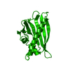
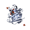
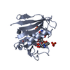
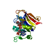
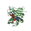
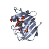
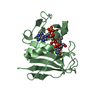
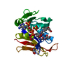
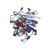
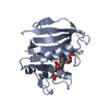
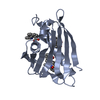
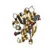
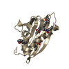
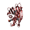
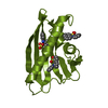
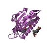
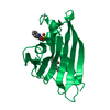
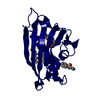
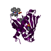
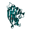
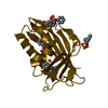
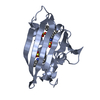
Details THE PROTEIN IS MONOMERIC IN SOLUTION, BUT FORMS DIMERS IN THE CRYSTAL STRUCTURE VIA ANTIPARALLEL INTERMOLECULAR BETA-SHEETS INVOLVING STRANDS BETA1. THE PAIRING SCHEME IS AB, CD, ..., YZ,...
 Movie
Movie Controller

 Yorodumi
 Open data
 Basic information
 Components
 Keywords
 Function and homology information
 Hypericum perforatum (plant)
 X-RAY DIFFRACTION /
  Authors
 Citation
 Structure visualization
 Downloads & links
 Other downloads


 PDBj
PDBj Assembly


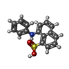
 Mass: 299.344 Da / Num. of mol.: 89 / Source method: obtained synthetically / Formula: C16H13NO3S
 Mass: 238.305 Da / Num. of mol.: 2 / Source method: obtained synthetically / Formula: C8H18N2O4S / Comment: pH buffer*YM
 Mass: 96.063 Da / Num. of mol.: 3 / Source method: obtained synthetically / Formula: SO4
 Mass: 18.015 Da / Num. of mol.: 35 / Source method: isolated from a natural source / Formula: H2O
 Sample preparation
 / Beamline: 22-BM / Wavelength: 1 Å
 Processing