 Movie
Movie Controller


+ Open data

- Basic information
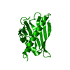
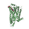
| Entry | Database: PDB / ID: 1bv1 | ||||||
|---|---|---|---|---|---|---|---|
| Title | BIRCH POLLEN ALLERGEN BET V 1 | ||||||
 Components Components | BET V 1 | ||||||
 Keywords Keywords | ALLERGEN / PATHOGENESIS-RELATED PROTEIN | ||||||
| Function / homology |  Function and homology information Function and homology informationabscisic acid binding / abscisic acid-activated signaling pathway / protein phosphatase inhibitor activity / defense response / signaling receptor activity / nucleus / cytoplasm Similarity search - Function | ||||||
| Biological species |  Betula pendula (European white birch) Betula pendula (European white birch) | ||||||
| Method |  X-RAY DIFFRACTION / MIR / Resolution: 2 Å X-RAY DIFFRACTION / MIR / Resolution: 2 Å | ||||||
 Authors Authors | Gajhede, M. / Osmark, P. / Poulsen, F.M. / Ipsen, H. / Larson, J.N. / Joostvan, R.J. / Schou, C. / Lowenstein, H. / Spangfort, M.D. | ||||||
 Citation Citation | Journal: Nat.Struct.Biol. / Year: 1996 Title: X-ray and NMR structure of Bet v 1, the origin of birch pollen allergy. Authors: Gajhede, M. / Osmark, P. / Poulsen, F.M. / Ipsen, H. / Larsen, J.N. / Joost van Neerven, R.J. / Schou, C. / Lowenstein, H. / Spangfort, M.D. | ||||||
| History |
|
- Structure visualization
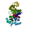
| Structure viewer | Molecule: MolmilJmol/JSmol |
|---|
- Downloads & links
-Download
| PDBx/mmCIF format | 1bv1.cif.gz | 44 KB | Display | PDBx/mmCIF format |
|---|---|---|---|---|
| PDB format | pdb1bv1.ent.gz | 31.7 KB | Display | PDB format |
| PDBx/mmJSON format | 1bv1.json.gz | Tree view | PDBx/mmJSON format | |
| Others |  Other downloads Other downloads |
-Validation report
| Arichive directory | https://data.pdbj.org/pub/pdb/validation_reports/bv/1bv1ftp://data.pdbj.org/pub/pdb/validation_reports/bv/1bv1 | HTTPS FTP |
|---|
-Related structure data


-Links
 PDBj
PDBj- Assembly
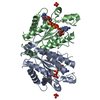
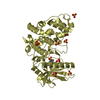
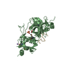
| Deposited unit | 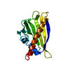
| ||||||||
|---|---|---|---|---|---|---|---|---|---|
| 1 | 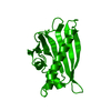
| ||||||||
| Unit cell |
|
-Components
| #1: Protein | Mass: 17427.576 Da / Num. of mol.: 1 Source method: isolated from a genetically manipulated source Source: (gene. exp.) Betula pendula (European white birch)Description: COMMERCIALLY AVAILABLE POLLEN FROM ALLERGEN, SWEDEN Cell: POLLEN / Cellular location: CYTOPLASM / Gene: BET V 1 1.2801 / Plasmid: PMALC / Cellular location (production host): CYTOPLASM / Gene (production host): MALE-BET V 1 / Production host:  |
|---|---|
| #2: Water | ChemComp-HOH /  Mass: 18.015 Da / Num. of mol.: 84 / Source method: isolated from a natural source / Formula: H2O Mass: 18.015 Da / Num. of mol.: 84 / Source method: isolated from a natural source / Formula: H2O |
-Experimental details
-Experiment
| Experiment | Method: X-RAY DIFFRACTION / Number of used crystals: 1 |
|---|
- Sample preparation
| Crystal | Density Matthews: 2.08 Å3/Da / Density % sol: 41 % | |||||||||||||||||||||||||||||||||||
|---|---|---|---|---|---|---|---|---|---|---|---|---|---|---|---|---|---|---|---|---|---|---|---|---|---|---|---|---|---|---|---|---|---|---|---|---|
| Crystal grow | pH: 6.3 Details: PROTEIN WAS CRYSTALLIZED BY MIXING 5 MG/ML PROTEIN SOLUTION WITH EQUAL AMOUNT OF 2.0 M AMS, 100 MM SODIUM CITRATE, PH 6.3 | |||||||||||||||||||||||||||||||||||
| Crystal grow | *PLUS Method: vapor diffusion, sitting drop / Details: seeding | |||||||||||||||||||||||||||||||||||
| Components of the solutions | *PLUS
|
-Data collection
| Diffraction | Mean temperature: 287 K |
|---|---|
| Diffraction source | Source: ROTATING ANODE / Type: RIGAKU RUH2R / Wavelength: 1.5418 |
| Detector | Type: RIGAKU / Detector: IMAGE PLATE / Date: Jan 1, 1996 |
| Radiation | Monochromator: GRAPHITE(002) / Monochromatic (M) / Laue (L): M / Scattering type: x-ray |
| Radiation wavelength | Wavelength: 1.5418 Å / Relative weight: 1 |
| Reflection | Resolution: 2→30 Å / Num. obs: 9554 / % possible obs: 95.8 % / Observed criterion σ(I): 0 / Redundancy: 4.8 % / Biso Wilson estimate: 14.6 Å2 / Rmerge(I) obs: 0.068 / Rsym value: 0.068 / Net I/σ(I): 14.6 |
| Reflection shell | Resolution: 2→2.03 Å / Redundancy: 3.2 % / Rmerge(I) obs: 0.36 / Mean I/σ(I) obs: 2.8 / Rsym value: 0.36 / % possible all: 84.2 |
| Reflection | *PLUS Num. measured all: 54458 |
| Reflection shell | *PLUS % possible obs: 84.2 % |
- Processing
| Software |
| ||||||||||||||||||||||||||||||||||||||||||||||||||||||||||||||||||||||||||||||||
|---|---|---|---|---|---|---|---|---|---|---|---|---|---|---|---|---|---|---|---|---|---|---|---|---|---|---|---|---|---|---|---|---|---|---|---|---|---|---|---|---|---|---|---|---|---|---|---|---|---|---|---|---|---|---|---|---|---|---|---|---|---|---|---|---|---|---|---|---|---|---|---|---|---|---|---|---|---|---|---|---|---|
| Refinement | Method to determine structure: MIR / Resolution: 2→40 Å / Rfactor Rfree error: 0.022 / Data cutoff high absF: 1000000 / Data cutoff low absF: 1.0E-5 / Isotropic thermal model: RESTRAINED / Cross valid method: THROUGHOUT / σ(F): 0 / Details: BULK SOLVENT CORRECTION
| ||||||||||||||||||||||||||||||||||||||||||||||||||||||||||||||||||||||||||||||||
| Displacement parameters | Biso mean: 25.2 Å2 | ||||||||||||||||||||||||||||||||||||||||||||||||||||||||||||||||||||||||||||||||
| Refine analyze | Luzzati coordinate error obs: 0.31 Å / Luzzati d res low obs: 40 Å | ||||||||||||||||||||||||||||||||||||||||||||||||||||||||||||||||||||||||||||||||
| Refinement step | Cycle: LAST / Resolution: 2→40 Å
| ||||||||||||||||||||||||||||||||||||||||||||||||||||||||||||||||||||||||||||||||
| Refine LS restraints |
| ||||||||||||||||||||||||||||||||||||||||||||||||||||||||||||||||||||||||||||||||
| LS refinement shell | Resolution: 2→2.09 Å / Total num. of bins used: 8
| ||||||||||||||||||||||||||||||||||||||||||||||||||||||||||||||||||||||||||||||||
| Xplor file |
| ||||||||||||||||||||||||||||||||||||||||||||||||||||||||||||||||||||||||||||||||
| Software | *PLUS Name: X-PLOR / Version: 3.851 / Classification: refinement | ||||||||||||||||||||||||||||||||||||||||||||||||||||||||||||||||||||||||||||||||
| Refinement | *PLUS | ||||||||||||||||||||||||||||||||||||||||||||||||||||||||||||||||||||||||||||||||
| Solvent computation | *PLUS | ||||||||||||||||||||||||||||||||||||||||||||||||||||||||||||||||||||||||||||||||
| Displacement parameters | *PLUS | ||||||||||||||||||||||||||||||||||||||||||||||||||||||||||||||||||||||||||||||||
| Refine LS restraints | *PLUS
| ||||||||||||||||||||||||||||||||||||||||||||||||||||||||||||||||||||||||||||||||
| LS refinement shell | *PLUS Rfactor obs: 0.263 |
