[English] 日本語
Yorodumi
- PDB-1qmr: BIRCH POLLEN ALLERGEN BET V 1 MUTANT N28T, K32Q, E45S, P108G -

+
Open data


ID or keywords:

Loading...

-
Basic information

Entry
Database: PDB / ID: 1qmr
TitleBIRCH POLLEN ALLERGEN BET V 1 MUTANT N28T, K32Q, E45S, P108G
ComponentsMAJOR POLLEN ALLERGEN BET V 1-A
KeywordsALLERGEN / PATHOGENESIS-RELATED PROTEIN
Function / homology
Function and homology information


response to biotic stimulus / abscisic acid binding / abscisic acid-activated signaling pathway / protein phosphatase inhibitor activity / defense response / signaling receptor activity / nucleus / cytoplasm
Similarity search - Function
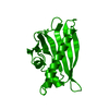
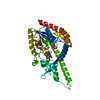
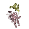
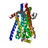
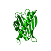
Pathogenesis-related protein Bet v I family / Pathogenesis-related proteins Bet v I family signature. / Bet v I type allergen / Bet v I/Major latex protein / Pathogenesis-related protein Bet v 1 family / START domain / Alpha-D-Glucose-1,6-Bisphosphate; Chain A, domain 4 / START-like domain superfamily / 2-Layer Sandwich / Alpha Beta
Similarity search - Domain/homology
Major pollen allergen Bet v 1-A
Similarity search - Component
Biological speciesBETULA VERRUCOSA (European white birch)
MethodX-RAY DIFFRACTION / MOLECULAR REPLACEMENT / Resolution: 2.15 Å
AuthorsHenriksen, A. / Holm, J.O. / Spangfort, M.D. / Gajhede, M.
Citation
Journal: J Immunol. / Year: 2004
Title: Allergy vaccine engineering: epitope modulation of recombinant Bet v 1 reduces IgE binding but retains protein folding pattern for induction of protective blocking-antibody responses.
Authors: Holm, J. / Gajhede, M. / Ferreras, M. / Henriksen, A. / Ipsen, H. / Larsen, J.N. / Lund, L. / Jacobi, H. / Millner, A. / Wurtzen, P.A. / Spangfort, M.D.
#1: Journal: Nat.Struct.Biol. / Year: 1996
Title: X-Ray and NMR Structure of Bet V 1, the Origin of Birch Pollen Allergy
Authors: Gajhede, M. / Osmark, P. / Poulsen, F.M. / Ipsen, H. / Larsen, J.N. / Van Neerven, R.J.J. / Schou, C. / Lowenstein, H. / Spangfort, M.D.
History
DepositionOct 6, 1999Deposition site: PDBE / Processing site: PDBE
Revision 1.0Oct 12, 2000Provider: repository / Type: Initial release
Revision 1.1May 8, 2011Group: Version format compliance
Revision 1.2Jul 13, 2011Group: Version format compliance
Revision 1.3Jul 5, 2017Group: Data collection / Category: diffrn_source / Item: _diffrn_source.type
Revision 1.4Oct 9, 2019Group: Data collection / Database references / Other / Category: citation / citation_author / pdbx_database_status
Item: _citation.journal_abbrev / _citation.journal_id_ASTM ..._citation.journal_abbrev / _citation.journal_id_ASTM / _citation.journal_id_CSD / _citation.page_last / _citation.pdbx_database_id_DOI / _citation.title / _citation_author.name / _pdbx_database_status.status_code_sf
Revision 1.5Dec 13, 2023Group: Data collection / Database references / Refinement description
Category: chem_comp_atom / chem_comp_bond ...chem_comp_atom / chem_comp_bond / database_2 / pdbx_initial_refinement_model
Item: _database_2.pdbx_DOI / _database_2.pdbx_database_accession

-
Structure visualization

Structure viewerMolecule:
MolmilJmol/JSmol

Downloads & links

-
Assembly

Deposited unit
A: MAJOR POLLEN ALLERGEN BET V 1-A


Theoretical massNumber of molelcules
Total (without water)17,3311
Polymers17,3311
Non-polymers00
Water82946
1
A: MAJOR POLLEN ALLERGEN BET V 1-A

A: MAJOR POLLEN ALLERGEN BET V 1-A


Theoretical massNumber of molelcules
Total (without water)34,6632
Polymers34,6632
Non-polymers00
Water362
TypeNameSymmetry operationNumber
identity operation1_555x,y,z1
crystal symmetry operation4_565x,-y+1,-z1
MethodPQS
Unit cell
Length a, b, c (Å)32.347, 74.196, 119.548
Angle α, β, γ (deg.)90.00, 90.00, 90.00
Int Tables number20
Space group name H-MC2221

-
Components

#1: Protein MAJOR POLLEN ALLERGEN BET V 1-A / BET V 1


Mass: 17331.428 Da / Num. of mol.: 1 / Mutation: YES
Source method: isolated from a genetically manipulated source
Source: (gene. exp.) BETULA VERRUCOSA (European white birch)
Cell: POLLEN / Cellular location: CYTOPLASM / Gene: BET V 1 1.2801 / Production host: PICHIA PASTORIS (fungus) / References: UniProt: P15494
#2: Water ChemComp-HOH / water


Mass: 18.015 Da / Num. of mol.: 46 / Source method: isolated from a natural source / Formula: H2O
Compound detailsCHAIN A ENGINEERED MUTATION ASN28THR, LYS32GLN, GLU45SER, PRO108GLY
Sequence detailsTHE LEU 62 SWS P15494 PHE 62 CONFLICT IS A KNOWN VARIANT FROM THE SWISSPROT SEQUENCE TAKEN FROM ...THE LEU 62 SWS P15494 PHE 62 CONFLICT IS A KNOWN VARIANT FROM THE SWISSPROT SEQUENCE TAKEN FROM REFERENCE: BREITENEDER H.,PETTENBURGER K.,BITO A.,VALENTA R., KRAFT D.,RUMPOLD H.,SCHEINER O.,BREITENBACH M. EMBO J. 8:1935-1938(1989).

-
Experimental details

-
Experiment

ExperimentMethod: X-RAY DIFFRACTION / Number of used crystals: 1

-
Sample preparation

CrystalDensity Matthews: 2.1 Å3/Da / Density % sol: 40.56 %
Crystal growpH: 6 / Details: pH 6.00
Crystal grow
*PLUS
Temperature: 25 ℃ / Method: vapor diffusion, sitting drop
Components of the solutions
*PLUS
IDConc.Common nameCrystal-IDSol-IDDetails
32.2 Mammonium sulfate1reservoir
4100 mMsodium citrate1reservoir
51 %dioxane1reservoir
60.01 %sodium azide1reservoir
1reservoir1drop
2rBet v 1 mutant1dropdialyzed against 10 mM Tris-HCl

-
Data collection

DiffractionMean temperature: 287 K
Diffraction sourceSource: ROTATING ANODE / Type: RIGAKU RUH2R / Wavelength: 1.5418
DetectorType: RIGAKU IMAGE PLATE / Detector: IMAGE PLATE / Date: Sep 15, 1999
RadiationProtocol: SINGLE WAVELENGTH / Monochromatic (M) / Laue (L): M / Scattering type: x-ray
Radiation wavelengthWavelength: 1.5418 Å / Relative weight: 1
ReflectionResolution: 2.15→37 Å / Num. obs: 7832 / % possible obs: 96.6 % / Redundancy: 5.6 % / Biso Wilson estimate: 11.4 Å2 / Rsym value: 0.04 / Net I/σ(I): 34
Reflection shellResolution: 2.15→2.23 Å / Redundancy: 3.7 % / Mean I/σ(I) obs: 9.5 / Rsym value: 0.135 / % possible all: 78.5
Reflection
*PLUS
Num. measured all: 55800 / Rmerge(I) obs: 0.04
Reflection shell
*PLUS
% possible obs: 78.5 % / Rmerge(I) obs: 0.165

-
Processing

Software
NameVersionClassification
CNS0.5refinement
DENZOdata reduction
SCALEPACKdata scaling
CNS0.5phasing
RefinementMethod to determine structure: MOLECULAR REPLACEMENT
Starting model: PDB ENTRY 1BV1
Resolution: 2.15→30 Å / Rfactor Rfree error: 0.012 / Data cutoff high absF: 248643.49 / Isotropic thermal model: RESTRAINED / Cross valid method: THROUGHOUT / σ(F): 0
RfactorNum. reflection% reflectionSelection details
Rfree0.238 407 5.2 %RANDOM
Rwork0.196 ---
obs0.196 7797 95.2 %-
Solvent computationSolvent model: FLAT MODEL / Bsol: 38.7618 Å2 / ksol: 0.330851 e/Å3
Displacement parametersBiso mean: 23.4 Å2
Baniso -1Baniso -2Baniso -3
1-0.47 Å20 Å20 Å2
2---4.5 Å20 Å2
3---4.03 Å2
Refine analyze
FreeObs
Luzzati coordinate error0.3 Å0.23 Å
Luzzati d res low-5 Å
Luzzati sigma a0.23 Å0.14 Å
Refinement stepCycle: LAST / Resolution: 2.15→30 Å
ProteinNucleic acidLigandSolventTotal
Num. atoms1223 0 0 46 1269
Refine LS restraints
Refine-IDTypeDev ideal
X-RAY DIFFRACTIONc_bond_d0.006
X-RAY DIFFRACTIONc_bond_d_na
X-RAY DIFFRACTIONc_bond_d_prot
X-RAY DIFFRACTIONc_angle_d
X-RAY DIFFRACTIONc_angle_d_na
X-RAY DIFFRACTIONc_angle_d_prot
X-RAY DIFFRACTIONc_angle_deg1.2
X-RAY DIFFRACTIONc_angle_deg_na
X-RAY DIFFRACTIONc_angle_deg_prot
X-RAY DIFFRACTIONc_dihedral_angle_d25
X-RAY DIFFRACTIONc_dihedral_angle_d_na
X-RAY DIFFRACTIONc_dihedral_angle_d_prot
X-RAY DIFFRACTIONc_improper_angle_d0.78
X-RAY DIFFRACTIONc_improper_angle_d_na
X-RAY DIFFRACTIONc_improper_angle_d_prot
X-RAY DIFFRACTIONc_mcbond_it
X-RAY DIFFRACTIONc_mcangle_it
X-RAY DIFFRACTIONc_scbond_it
X-RAY DIFFRACTIONc_scangle_it
LS refinement shellResolution: 2.15→2.28 Å / Rfactor Rfree error: 0.036 / Total num. of bins used: 6
RfactorNum. reflection% reflection
Rfree0.266 56 5.4 %
Rwork0.203 981 -
obs--78.1 %
Xplor file
Refine-IDSerial noParam fileTopol file
X-RAY DIFFRACTION1PROTEIN_REP.PARAMPROTEIN.TOP
X-RAY DIFFRACTION2WATER_REP.PARAMWATER.TOP
Software
*PLUS
Name: CNS / Version: 0.5 / Classification: refinement
Refine LS restraints
*PLUS
Refine-IDTypeDev ideal
X-RAY DIFFRACTIONc_dihedral_angle_d
X-RAY DIFFRACTIONc_dihedral_angle_deg25
X-RAY DIFFRACTIONc_improper_angle_d
X-RAY DIFFRACTIONc_improper_angle_deg0.78

+
About Yorodumi

-
News

-
Feb 9, 2022. New format data for meta-information of EMDB entries

New format data for meta-information of EMDB entries

  • Version 3 of the EMDB header file is now the official format.
  • The previous official version 1.9 will be removed from the archive.

Related info.:EMDB header

External links:wwPDB to switch to version 3 of the EMDB data model

-
Aug 12, 2020. Covid-19 info

Covid-19 info

URL: https://pdbjlvh1.pdbj.org/emnavi/covid19.php

New page: Covid-19 featured information page in EM Navigator.

Related info.:Covid-19 info / Mar 5, 2020. Novel coronavirus structure data

+
Mar 5, 2020. Novel coronavirus structure data

Novel coronavirus structure data

Related info.:Yorodumi Speices / Aug 12, 2020. Covid-19 info

External links:COVID-19 featured content - PDBj / Molecule of the Month (242):Coronavirus Proteases

+
Jan 31, 2019. EMDB accession codes are about to change! (news from PDBe EMDB page)

EMDB accession codes are about to change! (news from PDBe EMDB page)

  • The allocation of 4 digits for EMDB accession codes will soon come to an end. Whilst these codes will remain in use, new EMDB accession codes will include an additional digit and will expand incrementally as the available range of codes is exhausted. The current 4-digit format prefixed with “EMD-” (i.e. EMD-XXXX) will advance to a 5-digit format (i.e. EMD-XXXXX), and so on. It is currently estimated that the 4-digit codes will be depleted around Spring 2019, at which point the 5-digit format will come into force.
  • The EM Navigator/Yorodumi systems omit the EMD- prefix.

Related info.:Q: What is EMD? / ID/Accession-code notation in Yorodumi/EM Navigator

External links:EMDB Accession Codes are Changing Soon! / Contact to PDBj

+
Jul 12, 2017. Major update of PDB

Major update of PDB

  • wwPDB released updated PDB data conforming to the new PDBx/mmCIF dictionary.
  • This is a major update changing the version number from 4 to 5, and with Remediation, in which all the entries are updated.
  • In this update, many items about electron microscopy experimental information are reorganized (e.g. em_software).
  • Now, EM Navigator and Yorodumi are based on the updated data.

External links:wwPDB Remediation / Enriched Model Files Conforming to OneDep Data Standards Now Available in the PDB FTP Archive

-
Yorodumi

Thousand views of thousand structures

  • Yorodumi is a browser for structure data from EMDB, PDB, SASBDB, etc.
  • This page is also the successor to EM Navigator detail page, and also detail information page/front-end page for Omokage search.
  • The word "yorodu" (or yorozu) is an old Japanese word meaning "ten thousand". "mi" (miru) is to see.

Related info.:EMDB / PDB / SASBDB / Comparison of 3 databanks / Yorodumi Search / Aug 31, 2016. New EM Navigator & Yorodumi / Yorodumi Papers / Jmol/JSmol / Function and homology information / Changes in new EM Navigator and Yorodumi

Read more