 Movie
Movie Controller


+ Open data

- Basic information
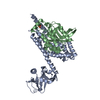
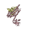
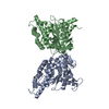
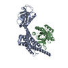
| Entry | Database: PDB / ID: 1jkw | ||||||
|---|---|---|---|---|---|---|---|
| Title | STRUCTURE OF CYCLIN MCS2 | ||||||
 Components Components | CYCLIN H | ||||||
 Keywords Keywords | CELL DIVISION / CYCLIN / CELL CYCLE / NUCLEAR PROTEIN | ||||||
| Function / homology |  Function and homology information Function and homology informationtranscription factor TFIIK complex / CAK-ERCC2 complex / transcription factor TFIIH core complex / transcription factor TFIIH holo complex / RNA Polymerase I Transcription Termination / cyclin-dependent protein serine/threonine kinase regulator activity / RNA Pol II CTD phosphorylation and interaction with CE during HIV infection / RNA Pol II CTD phosphorylation and interaction with CE / Formation of the Early Elongation Complex / Formation of the HIV-1 Early Elongation Complex ...transcription factor TFIIK complex / CAK-ERCC2 complex / transcription factor TFIIH core complex / transcription factor TFIIH holo complex / RNA Polymerase I Transcription Termination / cyclin-dependent protein serine/threonine kinase regulator activity / RNA Pol II CTD phosphorylation and interaction with CE during HIV infection / RNA Pol II CTD phosphorylation and interaction with CE / Formation of the Early Elongation Complex / Formation of the HIV-1 Early Elongation Complex / mRNA Capping / HIV Transcription Initiation / RNA Polymerase II HIV Promoter Escape / Transcription of the HIV genome / RNA Polymerase II Promoter Escape / RNA Polymerase II Transcription Pre-Initiation And Promoter Opening / RNA Polymerase II Transcription Initiation / RNA Polymerase II Transcription Initiation And Promoter Clearance / RNA Polymerase I Transcription Initiation / regulation of G1/S transition of mitotic cell cycle / Tat-mediated elongation of the HIV-1 transcript / Cyclin E associated events during G1/S transition / Formation of HIV-1 elongation complex containing HIV-1 Tat / Cyclin A:Cdk2-associated events at S phase entry / Formation of HIV elongation complex in the absence of HIV Tat / cyclin-dependent protein kinase holoenzyme complex / Cyclin A/B1/B2 associated events during G2/M transition / RNA Polymerase II Transcription Elongation / Formation of RNA Pol II elongation complex / RNA Polymerase II Pre-transcription Events / male germ cell nucleus / TP53 Regulates Transcription of DNA Repair Genes / transcription initiation at RNA polymerase II promoter / RNA Polymerase I Promoter Escape / NoRC negatively regulates rRNA expression / Transcription-Coupled Nucleotide Excision Repair (TC-NER) / Formation of TC-NER Pre-Incision Complex / Formation of Incision Complex in GG-NER / kinase activity / Cyclin D associated events in G1 / Dual incision in TC-NER / Gap-filling DNA repair synthesis and ligation in TC-NER / RUNX1 regulates transcription of genes involved in differentiation of HSCs / protein stabilization / regulation of transcription by RNA polymerase II / nucleoplasm / nucleus Similarity search - Function | ||||||
| Biological species |  Homo sapiens (human) Homo sapiens (human) | ||||||
| Method |  X-RAY DIFFRACTION / SYNCHROTRON / Resolution: 2.6 Å X-RAY DIFFRACTION / SYNCHROTRON / Resolution: 2.6 Å | ||||||
 Authors Authors | Andersen, G. / Poterszman, A. / Moras, D. / Thierry, J.C. | ||||||
 Citation Citation | Journal: FEBS Lett. / Year: 1996 Title: The crystal structure of human cyclin H. Authors: Andersen, G. / Poterszman, A. / Egly, J.M. / Moras, D. / Thierry, J.C. #1: Journal: Embo J. / Year: 1997 Title: The structure of cyclin H: common mode of kinase activation and specific features. Authors: Andersen, G. / Busso, D. / Poterszman, A. / Hwang, J.R. / Wurtz, J.M. / Ripp, R. / Thierry, J.C. / Egly, J.M. / Moras, D. #2: Journal: Structure / Year: 1995Title: The Crystal Structure of Cyclin A Authors: Brown, N.R. / Noble, M.E. / Endicott, J.A. / Garman, E.F. / Wakatsuki, S. / Mitchell, E. / Rasmussen, B. / Hunt, T. / Johnson, L.N. #3: Journal: Nature / Year: 1995Title: Mechanism of Cdk Activation Revealed by the Structure of a Cyclina-Cdk2 Complex Authors: Jeffrey, P.D. / Russo, A.A. / Polyak, K. / Gibbs, E. / Hurwitz, J. / Massague, J. / Pavletich, N.P. | ||||||
| History |
|
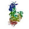
- Structure visualization
| Structure viewer | Molecule: MolmilJmol/JSmol |
|---|
- Downloads & links
-Download
| PDBx/mmCIF format | 1jkw.cif.gz | 69 KB | Display | PDBx/mmCIF format |
|---|---|---|---|---|
| PDB format | pdb1jkw.ent.gz | 51.6 KB | Display | PDB format |
| PDBx/mmJSON format | 1jkw.json.gz | Tree view | PDBx/mmJSON format | |
| Others |  Other downloads Other downloads |
-Validation report
| Arichive directory | https://data.pdbj.org/pub/pdb/validation_reports/jk/1jkwftp://data.pdbj.org/pub/pdb/validation_reports/jk/1jkw | HTTPS FTP |
|---|
-Related structure data
| Similar structure data |
|---|


-Links
 PDBj
PDBj


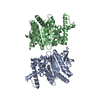
- Assembly
| Deposited unit | 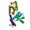
| ||||||||
|---|---|---|---|---|---|---|---|---|---|
| 1 | 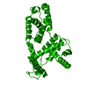
| ||||||||
| Unit cell |
|
-Components
| #1: Protein | Mass: 37681.445 Da / Num. of mol.: 1 Source method: isolated from a genetically manipulated source Source: (gene. exp.) Homo sapiens (human) / Description: HIS-TAG REMOVED BY THROMBIN DIGESTION / Gene: HUMAN CYCLIN H / Plasmid: PET15B / Gene (production host): HUMAN CYCLIN H / Production host:  |
|---|---|
| #2: Water | ChemComp-HOH /  Mass: 18.015 Da / Num. of mol.: 51 / Source method: isolated from a natural source / Formula: H2O Mass: 18.015 Da / Num. of mol.: 51 / Source method: isolated from a natural source / Formula: H2O |
| Has protein modification | N |
-Experimental details
-Experiment
| Experiment | Method: X-RAY DIFFRACTION |
|---|
- Sample preparation
| Crystal | Density Matthews: 4.37 Å3/Da / Density % sol: 72 % | |||||||||||||||||||||||||
|---|---|---|---|---|---|---|---|---|---|---|---|---|---|---|---|---|---|---|---|---|---|---|---|---|---|---|
| Crystal | *PLUS | |||||||||||||||||||||||||
| Crystal grow | *PLUS Temperature: 4 ℃ / pH: 5.8 / Method: vapor diffusion, hanging drop | |||||||||||||||||||||||||
| Components of the solutions | *PLUS
|
-Data collection
| Diffraction | Mean temperature: 120 K |
|---|---|
| Diffraction source | Source: SYNCHROTRON / Site: LURE  / Type: LURE / Wavelength: 0.91 / Type: LURE / Wavelength: 0.91 |
| Detector | Type: MARRESEARCH / Detector: IMAGE PLATE / Date: May 20, 1996 |
| Radiation | Monochromatic (M) / Laue (L): M / Scattering type: x-ray |
| Radiation wavelength | Wavelength: 0.91 Å / Relative weight: 1 |
| Reflection | Num. obs: 20809 / % possible obs: 98.5 % / Observed criterion σ(I): 0 / Redundancy: 2.8 % / Rmerge(I) obs: 0.059 |
| Reflection | *PLUS Highest resolution: 2.6 Å / Lowest resolution: 10 Å / Num. measured all: 21133 |
- Processing
| Software |
| ||||||||||||||||||||||||||||||||||||||||||||||||||||||||||||
|---|---|---|---|---|---|---|---|---|---|---|---|---|---|---|---|---|---|---|---|---|---|---|---|---|---|---|---|---|---|---|---|---|---|---|---|---|---|---|---|---|---|---|---|---|---|---|---|---|---|---|---|---|---|---|---|---|---|---|---|---|---|
| Refinement | Resolution: 2.6→14 Å / σ(F): 2 Details: THE LOOP LEU 236 - THR 243 HAS LIMITED DENSITY, AND IS NOT VERY RELIABLE.
| ||||||||||||||||||||||||||||||||||||||||||||||||||||||||||||
| Displacement parameters | Biso mean: 55.9 Å2 | ||||||||||||||||||||||||||||||||||||||||||||||||||||||||||||
| Refine analyze | Luzzati sigma a obs: 0.43 Å | ||||||||||||||||||||||||||||||||||||||||||||||||||||||||||||
| Refinement step | Cycle: LAST / Resolution: 2.6→14 Å
| ||||||||||||||||||||||||||||||||||||||||||||||||||||||||||||
| Refine LS restraints |
|
