 Movie
Movie Controller

[English] 日本語
 Yorodumi
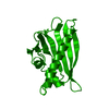
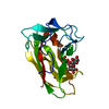
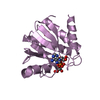
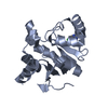
- PDB-2flh: Crystal structure of cytokinin-specific binding protein from mung... -
+ Open data

- Basic information
| Entry | Database: PDB / ID: 2flh | ||||||
|---|---|---|---|---|---|---|---|
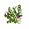
| Title | Crystal structure of cytokinin-specific binding protein from mung bean in complex with cytokinin | ||||||
 Components Components | cytokinin-specific binding protein | ||||||
 Keywords Keywords | PLANT PROTEIN / cytokinins / zeatin / pathogenesis-related proteins / multiple-ligand binding | ||||||
| Function / homology |  Function and homology information Function and homology informationcytokinin binding / gibberellin binding / abscisic acid binding / abscisic acid-activated signaling pathway / protein phosphatase inhibitor activity / defense response / signaling receptor activity / nucleus / cytoplasm Similarity search - Function | ||||||
| Biological species |  Vigna radiata (mung bean) Vigna radiata (mung bean) | ||||||
| Method |  X-RAY DIFFRACTION / SYNCHROTRON / MAD / Resolution: 1.2 Å X-RAY DIFFRACTION / SYNCHROTRON / MAD / Resolution: 1.2 Å | ||||||
 Authors Authors | Pasternak, O. / Bujacz, G.D. / Sikorski, M.M. / Jaskolski, M. | ||||||
 Citation Citation | Journal: Plant Cell / Year: 2006 Title: Crystal Structure of Vigna radiata Cytokinin-Specific Binding Protein in Complex with Zeatin. Authors: Pasternak, O. / Bujacz, G.D. / Fujimoto, Y. / Hashimoto, Y. / Jelen, F. / Otlewski, J. / Sikorski, M.M. / Jaskolski, M. #1: Journal: Acta Crystallogr.,Sect.D / Year: 2003 Title: Crystallization and preliminary crystallographic studies of mung bean cytokinin-specific binding protein Authors: Bujacz, G. / Pasternak, O. / Fujimoto, Y. / Hashimoto, Y. / Sikorski, M.M. / Jaskolski, M. #2: Journal: Eur.J.Biochem. / Year: 1998 Title: Purification and cDNA cloning of cytokinin-specific binding protein from mung bean (Vigna radiata) Authors: Fujimoto, Y. / Nagata, R. / Fukasawa, H. / Yano, K. / Azuma, M. / Iida, A. / Sugimoto, S. / Shudo, K. / Hashimoto, Y. #3: Journal: J.Mol.Biol. / Year: 2002Title: Crystal structures of two homologous pathogenesis-related proteins from yellow lupine Authors: Biesiadka, J. / Bujacz, G. / Sikorski, M.M. / Jaskolski, M. #4: Journal: Acta Crystallogr.,Sect.D / Year: 2005Title: Structure of a yellow lupine pathogenesis-related PR-10 protein belonging to a novel subclass Authors: Pasternak, O. / Biesiadka, J. / Dolot, R. / Handschuh, L. / Bujacz, G. / Sikorski, M.M. / Jaskolski, M. #5: Journal: Nat.Struct.Biol. / Year: 1996Title: X-ray and NMR structure of Bet v 1, the origin of birch pollen allergy. Authors: Gajhede, M. / Osmark, P. / Poulsen, F.M. / Ipsen, H. / Larsen, J.N. / van Neerven, R.J.J. / Schou, C. / Lowenstein, H. / Spangford, M.D. | ||||||
| History |
| ||||||
| Remark 600 | HETEROGEN The het group ZEA is also known as (E)-6-(4-HYDROXY-3- METHYL-BUT-2-ENYLAMINO)PURINE | ||||||
| Remark 999 | SEQUENCE According to authors, there is an error in the GB entry GB 4190976 at position 92 (Asn ...SEQUENCE According to authors, there is an error in the GB entry GB 4190976 at position 92 (Asn instead of Ser) |
- Structure visualization
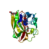
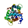
| Structure viewer | Molecule: MolmilJmol/JSmol |
|---|
- Downloads & links
-Download
| PDBx/mmCIF format | 2flh.cif.gz | 299 KB | Display | PDBx/mmCIF format |
|---|---|---|---|---|
| PDB format | pdb2flh.ent.gz | 242.6 KB | Display | PDB format |
| PDBx/mmJSON format | 2flh.json.gz | Tree view | PDBx/mmJSON format | |
| Others |  Other downloads Other downloads |
-Validation report
| Arichive directory | https://data.pdbj.org/pub/pdb/validation_reports/fl/2flhftp://data.pdbj.org/pub/pdb/validation_reports/fl/2flh | HTTPS FTP |
|---|
-Related structure data
| Related structure data | |
|---|---|
| Similar structure data |


-Links
 PDBj
PDBj- Assembly
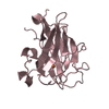
| Deposited unit | 
| ||||||||
|---|---|---|---|---|---|---|---|---|---|
| 1 | 
| ||||||||
| 2 | 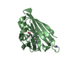
| ||||||||
| 3 | 
| ||||||||
| 4 | 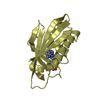
| ||||||||
| Unit cell |
| ||||||||
| Details | The biological assembly is a monomer. |
-Components
| #1: Protein | Mass: 17612.852 Da / Num. of mol.: 4 Source method: isolated from a genetically manipulated source Source: (gene. exp.) Vigna radiata (mung bean) / Gene: vrcsbp / Plasmid: pET-3a / Species (production host): Escherichia coli / Production host:  #2: Chemical | ChemComp-ZEA / (   Mass: 219.243 Da / Num. of mol.: 9 / Source method: obtained synthetically / Formula: C10H13N5O Mass: 219.243 Da / Num. of mol.: 9 / Source method: obtained synthetically / Formula: C10H13N5O#3: Chemical |   Mass: 22.990 Da / Num. of mol.: 2 / Source method: obtained synthetically / Formula: Na Mass: 22.990 Da / Num. of mol.: 2 / Source method: obtained synthetically / Formula: Na#4: Water | ChemComp-HOH / |  Mass: 18.015 Da / Num. of mol.: 645 / Source method: isolated from a natural source / Formula: H2O Mass: 18.015 Da / Num. of mol.: 645 / Source method: isolated from a natural source / Formula: H2O |
|---|
-Experimental details
-Experiment
| Experiment | Method: X-RAY DIFFRACTION / Number of used crystals: 1 |
|---|
- Sample preparation
| Crystal | Density Matthews: 2.31 Å3/Da / Density % sol: 46.68 % |
|---|---|
| Crystal grow | Temperature: 292 K / pH: 7.5 Details: sodium citrate, HEPES, zeatin, pH 7.5, VAPOR DIFFUSION, HANGING DROP, temperature 292K, pH 7.50 |
-Data collection
| Diffraction | Mean temperature: 100 K | |||||||||||||||
|---|---|---|---|---|---|---|---|---|---|---|---|---|---|---|---|---|
| Diffraction source | Source: SYNCHROTRON / Site: EMBL/DESY, HAMBURG  / Beamline: BW7B / Wavelength: 0.844, 1.2547, 1.2580, 1.2703 / Beamline: BW7B / Wavelength: 0.844, 1.2547, 1.2580, 1.2703 | |||||||||||||||
| Detector | Type: MARRESEARCH / Detector: IMAGE PLATE / Date: Aug 15, 2002 / Details: MIRRORS | |||||||||||||||
| Radiation | Monochromator: TRIANGULAR MONOCHROMATOR / Protocol: MAD / Monochromatic (M) / Laue (L): M / Scattering type: x-ray | |||||||||||||||
| Radiation wavelength |
| |||||||||||||||
| Reflection | Resolution: 1.2→30 Å / Num. obs: 189769 / % possible obs: 95.7 % / Observed criterion σ(I): -3 / Redundancy: 6.7 % / Biso Wilson estimate: 14.6 Å2 / Rmerge(I) obs: 0.07 / Net I/σ(I): 19.7 | |||||||||||||||
| Reflection shell | Resolution: 1.2→1.22 Å / Redundancy: 4.5 % / Rmerge(I) obs: 0.639 / Mean I/σ(I) obs: 2.6 / % possible all: 93.2 |
- Processing
| Software |
| |||||||||||||||||||||||||||||||||
|---|---|---|---|---|---|---|---|---|---|---|---|---|---|---|---|---|---|---|---|---|---|---|---|---|---|---|---|---|---|---|---|---|---|---|
| Refinement | Method to determine structure: MAD / Resolution: 1.2→15 Å / Cross valid method: THROUGHOUT / σ(F): 0 / Stereochemistry target values: Engh & Huber Details: ANISOTROPIC DISPLACEMENT PARAMETERS. H ATOMS AT RIDING POSITIONS. SEVERAL RESIDUES COULD NOT BE MODELED AT THE C-TERMINI (152-155 IN A, 154-155 IN B, 153-155 IN C, 155 IN A) AND IN ONE LOOP ...Details: ANISOTROPIC DISPLACEMENT PARAMETERS. H ATOMS AT RIDING POSITIONS. SEVERAL RESIDUES COULD NOT BE MODELED AT THE C-TERMINI (152-155 IN A, 154-155 IN B, 153-155 IN C, 155 IN A) AND IN ONE LOOP OF MOLECULE A (123-129). LONG SIDE CHAINS OF SOME SURFACE RESIDUES WERE POORLY VISIBLE IN ELECTRON DENSITY MAPS AND WERE MODELED WITH 0.0 OCCUPANCY
| |||||||||||||||||||||||||||||||||
| Solvent computation | Solvent model: BABINET MODEL | |||||||||||||||||||||||||||||||||
| Displacement parameters | Biso mean: 22.1 Å2 | |||||||||||||||||||||||||||||||||
| Refinement step | Cycle: LAST / Resolution: 1.2→15 Å
| |||||||||||||||||||||||||||||||||
| Refine LS restraints |
|
