 Movie
Movie Controller

[English] 日本語
 Yorodumi
- PDB-4dq6: Crystal structure of PLP-bound putative aminotransferase from Clo... -
+ Open data

- Basic information
| Entry | Database: PDB / ID: 4dq6 | ||||||
|---|---|---|---|---|---|---|---|
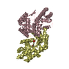
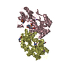
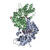
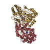
| Title | Crystal structure of PLP-bound putative aminotransferase from Clostridium difficile 630 | ||||||
 Components Components | Putative pyridoxal phosphate-dependent transferase | ||||||
 Keywords Keywords | TRANSFERASE / Structural Genomics / NIAID / National Institute of Allergy and Infectious Diseases / Center for Structural Genomics of Infectious Diseases / CSGID / internal aldimine with pyridoxalphosphate | ||||||
| Function / homology |  Function and homology information Function and homology informationcysteine-S-conjugate beta-lyase activity / cysteine-S-conjugate beta-lyase / pyridoxal phosphate binding / transferase activity / metal ion binding Similarity search - Function | ||||||
| Biological species |  Clostridium difficile (bacteria) Clostridium difficile (bacteria) | ||||||
| Method |  X-RAY DIFFRACTION / SYNCHROTRON / MOLECULAR REPLACEMENT / Resolution: 1.5 Å X-RAY DIFFRACTION / SYNCHROTRON / MOLECULAR REPLACEMENT / Resolution: 1.5 Å | ||||||
 Authors Authors | Shabalin, I.G. / Onopriyenko, O. / Kudritska, M. / Chruszcz, M. / Grimshaw, S. / Porebski, P.J. / Cooper, D.R. / Savchenko, A. / Anderson, W.F. / Minor, W. / Center for Structural Genomics of Infectious Diseases (CSGID) | ||||||
 Citation Citation | Journal: Microbiol Resour Announc / Year: 2023 Title: A high-throughput structural system biology approach to increase structure representation of proteins from Clostridioides difficile. Authors: Rosas-Lemus, M. / Dey, S. / Minasov, G. / Tan, K. / Anderson, S.M. / Brunzelle, J. / Nocadello, S. / Shabalin, I. / Filippova, E. / Halavaty, A. / Kim, Y. / Maltseva, N. / Osipiuk, J. / ...Authors: Rosas-Lemus, M. / Dey, S. / Minasov, G. / Tan, K. / Anderson, S.M. / Brunzelle, J. / Nocadello, S. / Shabalin, I. / Filippova, E. / Halavaty, A. / Kim, Y. / Maltseva, N. / Osipiuk, J. / Minor, W. / Joachimiak, A. / Savchenko, A. / Anderson, W.F. / Satchell, K.J.F. | ||||||
| History |
|
- Structure visualization
| Structure viewer | Molecule: MolmilJmol/JSmol |
|---|
- Downloads & links
-Download
| PDBx/mmCIF format | 4dq6.cif.gz | 340.4 KB | Display | PDBx/mmCIF format |
|---|---|---|---|---|
| PDB format | pdb4dq6.ent.gz | 276.4 KB | Display | PDB format |
| PDBx/mmJSON format | 4dq6.json.gz | Tree view | PDBx/mmJSON format | |
| Others |  Other downloads Other downloads |
-Validation report
| Arichive directory | https://data.pdbj.org/pub/pdb/validation_reports/dq/4dq6ftp://data.pdbj.org/pub/pdb/validation_reports/dq/4dq6 | HTTPS FTP |
|---|
-Related structure data
| Related structure data | 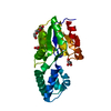 3sd7C  3srtC  3uuwC 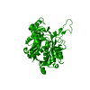 4dd5C  4dgtSC  4dunC 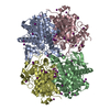 4e1lC 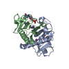 4eguC 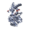 4gibC 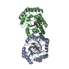 4h3dC  4isxC 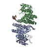 4jjpC 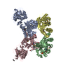 4kd5C  4mfgC  4nmyC  4rn7C 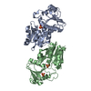 5dzsC  5ttaC 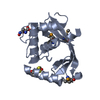 5tv7C 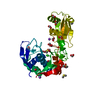 5txuC 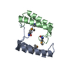 6n7mC  6ue2C  6wy4C  7k1uC  7rl8C  7rlrC S: Starting model for refinement C: citing same article ( |
|---|---|
| Similar structure data | |
| Other databases |


-Links
 PDBj
PDBj- Assembly
| Deposited unit | 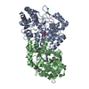
| ||||||||
|---|---|---|---|---|---|---|---|---|---|
| 1 |
| ||||||||
| Unit cell |
|
-Components
| #1: Protein | Mass: 45086.504 Da / Num. of mol.: 2 Source method: isolated from a genetically manipulated source Source: (gene. exp.) Clostridium difficile (bacteria) / Strain: 630 / Gene: CD2733, CD630_27330 / Plasmid: pMCSG7 / Production host: #2: Chemical |   Mass: 247.142 Da / Num. of mol.: 2 / Source method: obtained synthetically / Formula: C8H10NO6P Mass: 247.142 Da / Num. of mol.: 2 / Source method: obtained synthetically / Formula: C8H10NO6P#3: Chemical |   Mass: 35.453 Da / Num. of mol.: 3 / Source method: obtained synthetically / Formula: Cl Mass: 35.453 Da / Num. of mol.: 3 / Source method: obtained synthetically / Formula: Cl#4: Water | ChemComp-HOH / |  Mass: 18.015 Da / Num. of mol.: 1032 / Source method: isolated from a natural source / Formula: H2O Mass: 18.015 Da / Num. of mol.: 1032 / Source method: isolated from a natural source / Formula: H2OHas protein modification | N | |
|---|
-Experimental details
-Experiment
| Experiment | Method: X-RAY DIFFRACTION / Number of used crystals: 1 |
|---|
- Sample preparation
| Crystal | Density Matthews: 2.08 Å3/Da / Density % sol: 40.83 % |
|---|---|
| Crystal grow | Temperature: 293 K / Method: vapor diffusion, hanging drop / pH: 6 Details: Protein: 0.3M NaCl, 10mM Hepes pH 7.5. Precipitant: 25%PEG 2K MME, 0.2M Na Chloride, 0.1M Na Citrate, VAPOR DIFFUSION, HANGING DROP, temperature 293K |
-Data collection
| Diffraction | Mean temperature: 100 K | |||||||||||||||||||||||||||||||||||||||||||||||||||||||||||||||||||||||||||||||||||||||||||||||||||||||||||||||||||||||||||||||||||||||||||||||||||
|---|---|---|---|---|---|---|---|---|---|---|---|---|---|---|---|---|---|---|---|---|---|---|---|---|---|---|---|---|---|---|---|---|---|---|---|---|---|---|---|---|---|---|---|---|---|---|---|---|---|---|---|---|---|---|---|---|---|---|---|---|---|---|---|---|---|---|---|---|---|---|---|---|---|---|---|---|---|---|---|---|---|---|---|---|---|---|---|---|---|---|---|---|---|---|---|---|---|---|---|---|---|---|---|---|---|---|---|---|---|---|---|---|---|---|---|---|---|---|---|---|---|---|---|---|---|---|---|---|---|---|---|---|---|---|---|---|---|---|---|---|---|---|---|---|---|---|---|---|
| Diffraction source | Source: SYNCHROTRON / Site: APS  / Beamline: 19-ID / Wavelength: 0.97718 Å / Beamline: 19-ID / Wavelength: 0.97718 Å | |||||||||||||||||||||||||||||||||||||||||||||||||||||||||||||||||||||||||||||||||||||||||||||||||||||||||||||||||||||||||||||||||||||||||||||||||||
| Detector | Type: ADSC QUANTUM 315r / Detector: CCD / Date: Jul 6, 2011 / Details: mirrors | |||||||||||||||||||||||||||||||||||||||||||||||||||||||||||||||||||||||||||||||||||||||||||||||||||||||||||||||||||||||||||||||||||||||||||||||||||
| Radiation | Monochromator: Si 111 channel / Protocol: SINGLE WAVELENGTH / Monochromatic (M) / Laue (L): M / Scattering type: x-ray | |||||||||||||||||||||||||||||||||||||||||||||||||||||||||||||||||||||||||||||||||||||||||||||||||||||||||||||||||||||||||||||||||||||||||||||||||||
| Radiation wavelength | Wavelength: 0.97718 Å / Relative weight: 1 | |||||||||||||||||||||||||||||||||||||||||||||||||||||||||||||||||||||||||||||||||||||||||||||||||||||||||||||||||||||||||||||||||||||||||||||||||||
| Reflection | Resolution: 1.5→50 Å / Num. all: 119055 / Num. obs: 118460 / % possible obs: 99.5 % / Observed criterion σ(F): 0 / Observed criterion σ(I): 0 / Redundancy: 4 % / Biso Wilson estimate: 15.9 Å2 / Rmerge(I) obs: 0.072 / Rsym value: 0.072 / Χ2: 1.188 / Net I/σ(I): 21.5 | |||||||||||||||||||||||||||||||||||||||||||||||||||||||||||||||||||||||||||||||||||||||||||||||||||||||||||||||||||||||||||||||||||||||||||||||||||
| Reflection shell | Diffraction-ID: 1
|
- Processing
| Software |
| |||||||||||||||||||||||||||||||||||||||||||||||||||||||||||||||||||||||||||||||||||||||||||||||||||||||||||||||||||||||||||||||||||||||||||||||||||||||||||||||||||||||||||||||
|---|---|---|---|---|---|---|---|---|---|---|---|---|---|---|---|---|---|---|---|---|---|---|---|---|---|---|---|---|---|---|---|---|---|---|---|---|---|---|---|---|---|---|---|---|---|---|---|---|---|---|---|---|---|---|---|---|---|---|---|---|---|---|---|---|---|---|---|---|---|---|---|---|---|---|---|---|---|---|---|---|---|---|---|---|---|---|---|---|---|---|---|---|---|---|---|---|---|---|---|---|---|---|---|---|---|---|---|---|---|---|---|---|---|---|---|---|---|---|---|---|---|---|---|---|---|---|---|---|---|---|---|---|---|---|---|---|---|---|---|---|---|---|---|---|---|---|---|---|---|---|---|---|---|---|---|---|---|---|---|---|---|---|---|---|---|---|---|---|---|---|---|---|---|---|---|---|
| Refinement | Method to determine structure: MOLECULAR REPLACEMENT Starting model: pdb entry 4DGT Resolution: 1.5→50 Å / Cor.coef. Fo:Fc: 0.976 / Cor.coef. Fo:Fc free: 0.963 / WRfactor Rfree: 0.1863 / WRfactor Rwork: 0.1522 / Occupancy max: 1 / Occupancy min: 0.5 / FOM work R set: 0.8617 / SU B: 2.993 / SU ML: 0.056 / SU R Cruickshank DPI: 0.0698 / SU Rfree: 0.0731 / Isotropic thermal model: Isotropic / Cross valid method: THROUGHOUT / σ(F): 0 / σ(I): 0 / ESU R: 0.07 / ESU R Free: 0.073 / Stereochemistry target values: MAXIMUM LIKELIHOOD Details: U VALUES : WITH TLS ADDED HYDROGENS HAVE BEEN ADDED IN THE RIDING POSITIONS
| |||||||||||||||||||||||||||||||||||||||||||||||||||||||||||||||||||||||||||||||||||||||||||||||||||||||||||||||||||||||||||||||||||||||||||||||||||||||||||||||||||||||||||||||
| Solvent computation | Ion probe radii: 0.8 Å / Shrinkage radii: 0.8 Å / VDW probe radii: 1.2 Å / Solvent model: BABINET MODEL WITH MASK | |||||||||||||||||||||||||||||||||||||||||||||||||||||||||||||||||||||||||||||||||||||||||||||||||||||||||||||||||||||||||||||||||||||||||||||||||||||||||||||||||||||||||||||||
| Displacement parameters | Biso max: 63.63 Å2 / Biso mean: 22.3319 Å2 / Biso min: 10.94 Å2
| |||||||||||||||||||||||||||||||||||||||||||||||||||||||||||||||||||||||||||||||||||||||||||||||||||||||||||||||||||||||||||||||||||||||||||||||||||||||||||||||||||||||||||||||
| Refinement step | Cycle: LAST / Resolution: 1.5→50 Å
| |||||||||||||||||||||||||||||||||||||||||||||||||||||||||||||||||||||||||||||||||||||||||||||||||||||||||||||||||||||||||||||||||||||||||||||||||||||||||||||||||||||||||||||||
| Refine LS restraints |
| |||||||||||||||||||||||||||||||||||||||||||||||||||||||||||||||||||||||||||||||||||||||||||||||||||||||||||||||||||||||||||||||||||||||||||||||||||||||||||||||||||||||||||||||
| LS refinement shell | Resolution: 1.5→1.539 Å / Total num. of bins used: 20
| |||||||||||||||||||||||||||||||||||||||||||||||||||||||||||||||||||||||||||||||||||||||||||||||||||||||||||||||||||||||||||||||||||||||||||||||||||||||||||||||||||||||||||||||
| Refinement TLS params. | Method: refined / Refine-ID: X-RAY DIFFRACTION
| |||||||||||||||||||||||||||||||||||||||||||||||||||||||||||||||||||||||||||||||||||||||||||||||||||||||||||||||||||||||||||||||||||||||||||||||||||||||||||||||||||||||||||||||
| Refinement TLS group |
|
