 Movie
Movie Controller

[English] 日本語
 Yorodumi
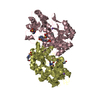
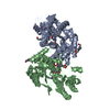
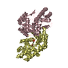
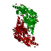
- PDB-3b1c: Crystal structure of betaC-S lyase from Streptococcus anginosus: ... -
+ Open data

- Basic information
| Entry | Database: PDB / ID: 3b1c | ||||||
|---|---|---|---|---|---|---|---|
| Title | Crystal structure of betaC-S lyase from Streptococcus anginosus: Internal aldimine form | ||||||
 Components Components | BetaC-S lyase | ||||||
 Keywords Keywords | LYASE | ||||||
| Function / homology |  Function and homology information Function and homology informationcysteine-S-conjugate beta-lyase activity / cysteine-S-conjugate beta-lyase / pyridoxal phosphate binding Similarity search - Function | ||||||
| Biological species |  Streptococcus anginosus (bacteria) Streptococcus anginosus (bacteria) | ||||||
| Method |  X-RAY DIFFRACTION / SYNCHROTRON / MOLECULAR REPLACEMENT / Resolution: 1.93 Å X-RAY DIFFRACTION / SYNCHROTRON / MOLECULAR REPLACEMENT / Resolution: 1.93 Å | ||||||
 Authors Authors | Kezuka, Y. / Yoshida, Y. / Nonaka, T. | ||||||
 Citation Citation | Journal: Proteins / Year: 2012 Title: Structural insights into catalysis by beta C-S lyase from Streptococcus anginosus Authors: Kezuka, Y. / Yoshida, Y. / Nonaka, T. #1: Journal: Acta Crystallogr.,Sect.F / Year: 2009 Title: Crystallization and preliminary X-ray analysis of betaC-S lyases from two oral Streptococci Authors: Kezuka, Y. / Yoshida, Y. / Nonaka, T. #2: Journal: Oral Microbiol.Immunol. / Year: 2008 Title: Molecular and enzymatic characterization of betaC-S lyase in Streptococcus constellatus Authors: Yoshida, Y. / Ito, S. / Sasaki, T. / Kishi, M. / Kurota, M. / Suwabe, A. / Kunimatsu, K. / Kato, H. #3: Journal: Biochem.Biophys.Res.Commun. / Year: 2003 Title: Differences in the betaC-S lyase activities of viridans group streptococci Authors: Yoshida, Y. / Negishi, M. / Amano, A. / Oho, T. / Nakano, Y. #4: Journal: Microbiology / Year: 2002 Title: lcd from Streptococcus anginosus encodes a C-S lyase with alpha,beta-elimination activity that degrades L-cysteine Authors: Yoshida, Y. / Nakano, Y. / Amano, A. / Yoshimura, M. / Fukamachi, H. / Oho, T. / Koga, T. | ||||||
| History |
|
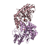
- Structure visualization
| Structure viewer | Molecule: MolmilJmol/JSmol |
|---|
- Downloads & links
-Download
| PDBx/mmCIF format | 3b1c.cif.gz | 354.2 KB | Display | PDBx/mmCIF format |
|---|---|---|---|---|
| PDB format | pdb3b1c.ent.gz | 287.5 KB | Display | PDB format |
| PDBx/mmJSON format | 3b1c.json.gz | Tree view | PDBx/mmJSON format | |
| Others |  Other downloads Other downloads |
-Validation report
| Arichive directory | https://data.pdbj.org/pub/pdb/validation_reports/b1/3b1cftp://data.pdbj.org/pub/pdb/validation_reports/b1/3b1c | HTTPS FTP |
|---|
-Related structure data


-Links
 PDBj
PDBj- Assembly
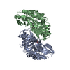
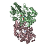
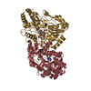
| Deposited unit | 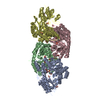
| ||||||||
|---|---|---|---|---|---|---|---|---|---|
| 1 | 
| ||||||||
| 2 | 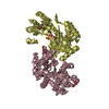
| ||||||||
| Unit cell |
|
-Components
| #1: Protein | Mass: 44649.309 Da / Num. of mol.: 4 Source method: isolated from a genetically manipulated source Source: (gene. exp.) Streptococcus anginosus (bacteria) / Strain: IMU102 / Gene: lcd / Plasmid: pGEX-6P-1 / Production host: #2: Chemical | ChemComp-PLP /   Mass: 247.142 Da / Num. of mol.: 4 / Source method: obtained synthetically / Formula: C8H10NO6P Mass: 247.142 Da / Num. of mol.: 4 / Source method: obtained synthetically / Formula: C8H10NO6P#3: Chemical | ChemComp-SO4 /   Mass: 96.063 Da / Num. of mol.: 6 / Source method: obtained synthetically / Formula: SO4 Mass: 96.063 Da / Num. of mol.: 6 / Source method: obtained synthetically / Formula: SO4#4: Chemical | ChemComp-GOL /   Mass: 92.094 Da / Num. of mol.: 14 / Source method: obtained synthetically / Formula: C3H8O3 Mass: 92.094 Da / Num. of mol.: 14 / Source method: obtained synthetically / Formula: C3H8O3#5: Water | ChemComp-HOH / |  Mass: 18.015 Da / Num. of mol.: 1466 / Source method: isolated from a natural source / Formula: H2O Mass: 18.015 Da / Num. of mol.: 1466 / Source method: isolated from a natural source / Formula: H2OHas protein modification | Y | |
|---|
-Experimental details
-Experiment
| Experiment | Method: X-RAY DIFFRACTION / Number of used crystals: 1 |
|---|
- Sample preparation
| Crystal | Density Matthews: 2.26 Å3/Da / Density % sol: 45.45 % |
|---|---|
| Crystal grow | Temperature: 293 K / Method: vapor diffusion, hanging drop / pH: 7.5 Details: 1.6M Ammonium sulfate, 0.1M Hepes pH7.5, 0.1M Sodium chloride, VAPOR DIFFUSION, HANGING DROP, temperature 293K |
-Data collection
| Diffraction | Mean temperature: 100 K |
|---|---|
| Diffraction source | Source: SYNCHROTRON / Site: SPring-8  / Beamline: BL38B1 / Wavelength: 1 Å / Beamline: BL38B1 / Wavelength: 1 Å |
| Detector | Type: RIGAKU JUPITER 210 / Detector: CCD / Date: Jul 26, 2008 |
| Radiation | Monochromator: Fixed exit Si (111) double crystal monochromator Protocol: SINGLE WAVELENGTH / Monochromatic (M) / Laue (L): M / Scattering type: x-ray |
| Radiation wavelength | Wavelength: 1 Å / Relative weight: 1 |
| Reflection | Resolution: 1.93→108.47 Å / Num. all: 122200 / Num. obs: 122200 / % possible obs: 100 % / Observed criterion σ(F): 0 / Observed criterion σ(I): 0 / Rmerge(I) obs: 0.061 / Rsym value: 0.061 |
| Reflection shell | Resolution: 1.93→1.97 Å / Rmerge(I) obs: 0.23 / Num. unique all: 7249 / Rsym value: 0.23 / % possible all: 100 |
- Processing
| Software |
| ||||||||||||||||||||||||||||||||||||||||||||||||||||||||||||||||||||||||||||||||||||||||||||||||||||||||||||||||||||||||||||||||||||||||||||||||||||||||||||||||||||||||||
|---|---|---|---|---|---|---|---|---|---|---|---|---|---|---|---|---|---|---|---|---|---|---|---|---|---|---|---|---|---|---|---|---|---|---|---|---|---|---|---|---|---|---|---|---|---|---|---|---|---|---|---|---|---|---|---|---|---|---|---|---|---|---|---|---|---|---|---|---|---|---|---|---|---|---|---|---|---|---|---|---|---|---|---|---|---|---|---|---|---|---|---|---|---|---|---|---|---|---|---|---|---|---|---|---|---|---|---|---|---|---|---|---|---|---|---|---|---|---|---|---|---|---|---|---|---|---|---|---|---|---|---|---|---|---|---|---|---|---|---|---|---|---|---|---|---|---|---|---|---|---|---|---|---|---|---|---|---|---|---|---|---|---|---|---|---|---|---|---|---|---|---|
| Refinement | Method to determine structure: MOLECULAR REPLACEMENT / Resolution: 1.93→108.47 Å / Cor.coef. Fo:Fc: 0.958 / Cor.coef. Fo:Fc free: 0.937 / SU B: 2.679 / SU ML: 0.081 / Cross valid method: THROUGHOUT / σ(I): 0 / ESU R Free: 0.13 / Stereochemistry target values: MAXIMUM LIKELIHOOD / Details: HYDROGENS HAVE BEEN ADDED IN THE RIDING POSITIONS
| ||||||||||||||||||||||||||||||||||||||||||||||||||||||||||||||||||||||||||||||||||||||||||||||||||||||||||||||||||||||||||||||||||||||||||||||||||||||||||||||||||||||||||
| Solvent computation | Ion probe radii: 0.8 Å / Shrinkage radii: 0.8 Å / VDW probe radii: 1.2 Å / Solvent model: MASK | ||||||||||||||||||||||||||||||||||||||||||||||||||||||||||||||||||||||||||||||||||||||||||||||||||||||||||||||||||||||||||||||||||||||||||||||||||||||||||||||||||||||||||
| Displacement parameters | Biso mean: 10.729 Å2
| ||||||||||||||||||||||||||||||||||||||||||||||||||||||||||||||||||||||||||||||||||||||||||||||||||||||||||||||||||||||||||||||||||||||||||||||||||||||||||||||||||||||||||
| Refinement step | Cycle: LAST / Resolution: 1.93→108.47 Å
| ||||||||||||||||||||||||||||||||||||||||||||||||||||||||||||||||||||||||||||||||||||||||||||||||||||||||||||||||||||||||||||||||||||||||||||||||||||||||||||||||||||||||||
| Refine LS restraints |
| ||||||||||||||||||||||||||||||||||||||||||||||||||||||||||||||||||||||||||||||||||||||||||||||||||||||||||||||||||||||||||||||||||||||||||||||||||||||||||||||||||||||||||
| LS refinement shell | Resolution: 1.93→1.98 Å / Total num. of bins used: 20
|
