 Movie
Movie Controller

[English] 日本語
 Yorodumi
- PDB-6qp3: Crystal structure of the PLP-bound C-S lyase from Bacillus subtil... -
+ Open data

- Basic information
| Entry | Database: PDB / ID: 6qp3 | ||||||
|---|---|---|---|---|---|---|---|
| Title | Crystal structure of the PLP-bound C-S lyase from Bacillus subtilis (strain 168) | ||||||
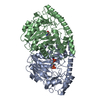
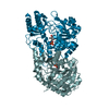
 Components Components | Cystathionine beta-lyase PatB | ||||||
 Keywords Keywords | LYASE / pyridoxal phosphate binding / Cysteine-S-conjugate beta-lyase | ||||||
| Function / homology |  Function and homology information Function and homology informationcysteine-S-conjugate beta-lyase activity / cysteine-S-conjugate beta-lyase / : / pyridoxal phosphate binding Similarity search - Function | ||||||
| Biological species |  | ||||||
| Method |  X-RAY DIFFRACTION / SYNCHROTRON / MOLECULAR REPLACEMENT / Resolution: 2.3 Å X-RAY DIFFRACTION / SYNCHROTRON / MOLECULAR REPLACEMENT / Resolution: 2.3 Å | ||||||
 Authors Authors | Herman, R. / Rudden, M. / Wilkinson, A.J. / Hanai, S. / Thomas, G.H. | ||||||
| Funding support |  United Kingdom, 1items United Kingdom, 1items
| ||||||
 Citation Citation | Journal: Sci Rep / Year: 2020 Title: The molecular basis of thioalcohol production in human body odour. Authors: Rudden, M. / Herman, R. / Rose, M. / Bawdon, D. / Cox, D.S. / Dodson, E. / Holden, M.T.G. / Wilkinson, A.J. / James, A.G. / Thomas, G.H. | ||||||
| History |
|
- Structure visualization
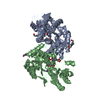
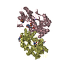
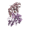
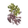
| Structure viewer | Molecule: MolmilJmol/JSmol |
|---|
- Downloads & links
-Download
| PDBx/mmCIF format | 6qp3.cif.gz | 299.6 KB | Display | PDBx/mmCIF format |
|---|---|---|---|---|
| PDB format | pdb6qp3.ent.gz | 243.9 KB | Display | PDB format |
| PDBx/mmJSON format | 6qp3.json.gz | Tree view | PDBx/mmJSON format | |
| Others |  Other downloads Other downloads |
-Validation report
| Arichive directory | https://data.pdbj.org/pub/pdb/validation_reports/qp/6qp3ftp://data.pdbj.org/pub/pdb/validation_reports/qp/6qp3 | HTTPS FTP |
|---|
-Related structure data
| Related structure data |  6qp1C  6qp2C  4dq6S  6rvi 6rvj C: citing same article ( S: Starting model for refinement |
|---|---|
| Similar structure data |


-Links
 PDBj
PDBj- Assembly
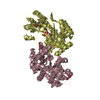
| Deposited unit | 
| ||||||||
|---|---|---|---|---|---|---|---|---|---|
| 1 | 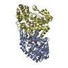
| ||||||||
| 2 | 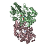
| ||||||||
| Unit cell |
|
-Components
| #1: Protein | Mass: 42838.805 Da / Num. of mol.: 4 Source method: isolated from a genetically manipulated source Source: (gene. exp.) Strain: 168 / Gene: patB, BSU31440 / Production host: References: UniProt: Q08432, cysteine-S-conjugate beta-lyase #2: Chemical | ChemComp-ACT /   Mass: 59.044 Da / Num. of mol.: 4 / Source method: obtained synthetically / Formula: C2H3O2 / Feature type: SUBJECT OF INVESTIGATION Mass: 59.044 Da / Num. of mol.: 4 / Source method: obtained synthetically / Formula: C2H3O2 / Feature type: SUBJECT OF INVESTIGATION#3: Chemical | ChemComp-PLP /   Mass: 247.142 Da / Num. of mol.: 4 / Source method: obtained synthetically / Formula: C8H10NO6P Mass: 247.142 Da / Num. of mol.: 4 / Source method: obtained synthetically / Formula: C8H10NO6P#4: Water | ChemComp-HOH / |  Mass: 18.015 Da / Num. of mol.: 84 / Source method: isolated from a natural source / Formula: H2O Mass: 18.015 Da / Num. of mol.: 84 / Source method: isolated from a natural source / Formula: H2O |
|---|
-Experimental details
-Experiment
| Experiment | Method: X-RAY DIFFRACTION / Number of used crystals: 1 |
|---|
- Sample preparation
| Crystal | Density Matthews: 2.42 Å3/Da / Density % sol: 49.09 % |
|---|---|
| Crystal grow | Temperature: 298.15 K / Method: vapor diffusion, sitting drop / pH: 7.1 / Details: 0.2M Ammonium acetate, 20% PEG 3,350, pH 7.1 / Temp details: Room temperature |
-Data collection
| Diffraction | Mean temperature: 120 K / Serial crystal experiment: N |
|---|---|
| Diffraction source | Source: SYNCHROTRON / Site: Diamond / Beamline: I04 / Wavelength: 0.9795 Å |
| Detector | Type: DECTRIS PILATUS 6M-F / Detector: PIXEL / Date: Oct 18, 2018 |
| Radiation | Protocol: SINGLE WAVELENGTH / Monochromatic (M) / Laue (L): M / Scattering type: x-ray |
| Radiation wavelength | Wavelength: 0.9795 Å / Relative weight: 1 |
| Reflection | Resolution: 2.3→61.28 Å / Num. obs: 72159 / % possible obs: 96.1 % / Redundancy: 7.9 % / CC1/2: 0.991 / Rmerge(I) obs: 0.197 / Rpim(I) all: 0.071 / Rrim(I) all: 0.21 / Net I/σ(I): 8.7 |
| Reflection shell | Resolution: 2.3→2.35 Å / Redundancy: 5.6 % / Rmerge(I) obs: 1.195 / Num. unique obs: 3803 / CC1/2: 0.574 / Rpim(I) all: 0.479 / Rrim(I) all: 1.295 / % possible all: 82.8 |
- Processing
| Software |
| ||||||||||||||||||||||||||||||||||||||||||||||||||||||||||||
|---|---|---|---|---|---|---|---|---|---|---|---|---|---|---|---|---|---|---|---|---|---|---|---|---|---|---|---|---|---|---|---|---|---|---|---|---|---|---|---|---|---|---|---|---|---|---|---|---|---|---|---|---|---|---|---|---|---|---|---|---|---|
| Refinement | Method to determine structure: MOLECULAR REPLACEMENT Starting model: 4DQ6 Resolution: 2.3→61.28 Å / Cor.coef. Fo:Fc: 0.937 / Cor.coef. Fo:Fc free: 0.894 / SU B: 10.48 / SU ML: 0.236 / Cross valid method: THROUGHOUT / σ(F): 0 / ESU R: 0.387 / ESU R Free: 0.257 Details: HYDROGENS HAVE BEEN ADDED IN THE RIDING POSITIONS U VALUES : REFINED INDIVIDUALLY
| ||||||||||||||||||||||||||||||||||||||||||||||||||||||||||||
| Solvent computation | Ion probe radii: 0.8 Å / Shrinkage radii: 0.8 Å / VDW probe radii: 1.2 Å | ||||||||||||||||||||||||||||||||||||||||||||||||||||||||||||
| Displacement parameters | Biso max: 124.43 Å2 / Biso mean: 27.599 Å2 / Biso min: 11.45 Å2
| ||||||||||||||||||||||||||||||||||||||||||||||||||||||||||||
| Refinement step | Cycle: final / Resolution: 2.3→61.28 Å
| ||||||||||||||||||||||||||||||||||||||||||||||||||||||||||||
| Refine LS restraints |
| ||||||||||||||||||||||||||||||||||||||||||||||||||||||||||||
| LS refinement shell | Resolution: 2.3→2.36 Å / Rfactor Rfree error: 0 / Total num. of bins used: 20
|
