 Movie
Movie Controller

[English] 日本語
 Yorodumi
- PDB-6qp1: Crystal structure of the PLP-bound C-S lyase in the external aldi... -
+ Open data

- Basic information
| Entry | Database: PDB / ID: 6qp1 | ||||||
|---|---|---|---|---|---|---|---|
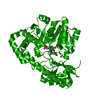
| Title | Crystal structure of the PLP-bound C-S lyase in the external aldimine form from Staphylococcus hominis complexed with an inhibitor, L-cycloserine. | ||||||
 Components Components | Aminotransferase | ||||||
 Keywords Keywords | LYASE / pyridoxal phosphate binding / cystathionine beta-lyase / inhibitor / complex / L-cycloserine / external aldimine | ||||||
| Function / homology |  Function and homology information Function and homology information | ||||||
| Biological species |  Staphylococcus hominis (bacteria) Staphylococcus hominis (bacteria) | ||||||
| Method |  X-RAY DIFFRACTION / SYNCHROTRON / MOLECULAR REPLACEMENT / Resolution: 1.42 Å X-RAY DIFFRACTION / SYNCHROTRON / MOLECULAR REPLACEMENT / Resolution: 1.42 Å | ||||||
 Authors Authors | Herman, R. / Rudden, M. / Wilkinson, A.J. / Hanai, S. / Thomas, G.H. | ||||||
| Funding support |  United Kingdom, 1items United Kingdom, 1items
| ||||||
 Citation Citation | Journal: Sci Rep / Year: 2020 Title: The molecular basis of thioalcohol production in human body odour. Authors: Rudden, M. / Herman, R. / Rose, M. / Bawdon, D. / Cox, D.S. / Dodson, E. / Holden, M.T.G. / Wilkinson, A.J. / James, A.G. / Thomas, G.H. | ||||||
| History |
|
- Structure visualization
| Structure viewer | Molecule: MolmilJmol/JSmol |
|---|
- Downloads & links
-Download
| PDBx/mmCIF format | 6qp1.cif.gz | 179.9 KB | Display | PDBx/mmCIF format |
|---|---|---|---|---|
| PDB format | pdb6qp1.ent.gz | 140.9 KB | Display | PDB format |
| PDBx/mmJSON format | 6qp1.json.gz | Tree view | PDBx/mmJSON format | |
| Others |  Other downloads Other downloads |
-Validation report
| Arichive directory | https://data.pdbj.org/pub/pdb/validation_reports/qp/6qp1ftp://data.pdbj.org/pub/pdb/validation_reports/qp/6qp1 | HTTPS FTP |
|---|
-Related structure data
| Related structure data |  6qp2C  6qp3C  4dq6S  6rvi 6rvj C: citing same article ( S: Starting model for refinement |
|---|---|
| Similar structure data |


-Links
 PDBj
PDBj- Assembly
| Deposited unit | 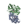
| ||||||||
|---|---|---|---|---|---|---|---|---|---|
| 1 |
| ||||||||
| Unit cell |
|
-Components
| #1: Protein | Mass: 48428.652 Da / Num. of mol.: 2 Source method: isolated from a genetically manipulated source Details: This sequence contains additional residues introduced by the expression vector (MGGGFA.....ENLYFQGHHHHHHHHHH) Source: (gene. exp.) Staphylococcus hominis (bacteria) / Gene: BUZ46_10400 / Plasmid: pBADcLIC / Production host: #2: Chemical |   Mass: 331.219 Da / Num. of mol.: 2 / Source method: obtained synthetically / Formula: C11H14N3O7P / Feature type: SUBJECT OF INVESTIGATION Mass: 331.219 Da / Num. of mol.: 2 / Source method: obtained synthetically / Formula: C11H14N3O7P / Feature type: SUBJECT OF INVESTIGATION#3: Water | ChemComp-HOH / |  Mass: 18.015 Da / Num. of mol.: 388 / Source method: isolated from a natural source / Formula: H2O Mass: 18.015 Da / Num. of mol.: 388 / Source method: isolated from a natural source / Formula: H2O |
|---|
-Experimental details
-Experiment
| Experiment | Method: X-RAY DIFFRACTION / Number of used crystals: 1 |
|---|
- Sample preparation
| Crystal | Density Matthews: 1.97 Å3/Da / Density % sol: 37.45 % |
|---|---|
| Crystal grow | Temperature: 298.15 K / Method: vapor diffusion, hanging drop / pH: 6 Details: 4% Tacsimate pH 6.0, 12% PEG 3,350 (F1: PEG/ION HT, Hampton Research), 20mM L-cycloserine |
-Data collection
| Diffraction | Mean temperature: 120 K / Serial crystal experiment: N |
|---|---|
| Diffraction source | Source: SYNCHROTRON / Site: Diamond / Beamline: I03 / Wavelength: 0.9762 Å |
| Detector | Type: DECTRIS PILATUS3 6M / Detector: PIXEL / Date: Jul 23, 2018 |
| Radiation | Monochromator: 0.9762 / Protocol: SINGLE WAVELENGTH / Monochromatic (M) / Laue (L): M / Scattering type: x-ray |
| Radiation wavelength | Wavelength: 0.9762 Å / Relative weight: 1 |
| Reflection | Resolution: 1.42→52.88 Å / Num. obs: 143873 / % possible obs: 99.6 % / Redundancy: 7.9 % / CC1/2: 0.997 / Rmerge(I) obs: 0.103 / Rpim(I) all: 0.039 / Rrim(I) all: 0.11 / Net I/σ(I): 9.8 |
| Reflection shell | Resolution: 1.42→1.44 Å / Redundancy: 8 % / Rmerge(I) obs: 2.503 / Num. unique obs: 7011 / CC1/2: 0.331 / Rpim(I) all: 0.929 / Rrim(I) all: 2.673 / % possible all: 99.1 |
- Processing
| Software |
| ||||||||||||||||||||||||||||||||||||||||||||||||||||||||||||
|---|---|---|---|---|---|---|---|---|---|---|---|---|---|---|---|---|---|---|---|---|---|---|---|---|---|---|---|---|---|---|---|---|---|---|---|---|---|---|---|---|---|---|---|---|---|---|---|---|---|---|---|---|---|---|---|---|---|---|---|---|---|
| Refinement | Method to determine structure: MOLECULAR REPLACEMENT Starting model: 4DQ6 Resolution: 1.42→51.99 Å / Cor.coef. Fo:Fc: 0.964 / Cor.coef. Fo:Fc free: 0.956 / SU B: 1.577 / SU ML: 0.059 / Cross valid method: THROUGHOUT / σ(F): 0 / ESU R: 0.07 / ESU R Free: 0.071 Details: HYDROGENS HAVE BEEN ADDED IN THE RIDING POSITIONS U VALUES : REFINED INDIVIDUALLY
| ||||||||||||||||||||||||||||||||||||||||||||||||||||||||||||
| Solvent computation | Ion probe radii: 0.8 Å / Shrinkage radii: 0.8 Å / VDW probe radii: 1.2 Å | ||||||||||||||||||||||||||||||||||||||||||||||||||||||||||||
| Displacement parameters | Biso max: 394.56 Å2 / Biso mean: 22.723 Å2 / Biso min: 10.64 Å2
| ||||||||||||||||||||||||||||||||||||||||||||||||||||||||||||
| Refinement step | Cycle: final / Resolution: 1.42→51.99 Å
| ||||||||||||||||||||||||||||||||||||||||||||||||||||||||||||
| Refine LS restraints |
| ||||||||||||||||||||||||||||||||||||||||||||||||||||||||||||
| LS refinement shell | Resolution: 1.42→1.457 Å / Rfactor Rfree error: 0 / Total num. of bins used: 20
|
