 Movie
Movie Controller

[English] 日本語
 Yorodumi
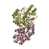
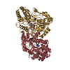
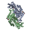
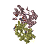
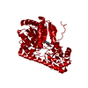
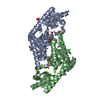
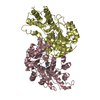
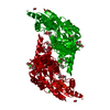
- PDB-1c7n: CRYSTAL STRUCTURE OF CYSTALYSIN FROM TREPONEMA DENTICOLA CONTAINS... -
+ Open data

- Basic information
| Entry | Database: PDB / ID: 1c7n | ||||||
|---|---|---|---|---|---|---|---|
| Title | CRYSTAL STRUCTURE OF CYSTALYSIN FROM TREPONEMA DENTICOLA CONTAINS A PYRIDOXAL 5'-PHOSPHATE COFACTOR | ||||||
 Components Components | CYSTALYSIN | ||||||
 Keywords Keywords | TRANSFERASE / Aminotransferase / Pyridoxal phosphate | ||||||
| Function / homology |  Function and homology information Function and homology informationcysteine-S-conjugate beta-lyase activity / cysteine-S-conjugate beta-lyase / pyridoxal phosphate binding Similarity search - Function | ||||||
| Biological species |  Treponema denticola (bacteria) Treponema denticola (bacteria) | ||||||
| Method |  X-RAY DIFFRACTION / SYNCHROTRON / Resolution: 1.9 Å X-RAY DIFFRACTION / SYNCHROTRON / Resolution: 1.9 Å | ||||||
 Authors Authors | Krupka, H.I. / Huber, R. / Holt, S.C. / Clausen, T. | ||||||
 Citation Citation | Journal: EMBO J. / Year: 2000 Title: Crystal structure of cystalysin from Treponema denticola: a pyridoxal 5'-phosphate-dependent protein acting as a haemolytic enzyme. Authors: Krupka, H.I. / Huber, R. / Holt, S.C. / Clausen, T. | ||||||
| History |
|
- Structure visualization
| Structure viewer | Molecule: MolmilJmol/JSmol |
|---|
- Downloads & links
-Download
| PDBx/mmCIF format | 1c7n.cif.gz | 675.5 KB | Display | PDBx/mmCIF format |
|---|---|---|---|---|
| PDB format | pdb1c7n.ent.gz | 559.4 KB | Display | PDB format |
| PDBx/mmJSON format | 1c7n.json.gz | Tree view | PDBx/mmJSON format | |
| Others |  Other downloads Other downloads |
-Validation report
| Arichive directory | https://data.pdbj.org/pub/pdb/validation_reports/c7/1c7nftp://data.pdbj.org/pub/pdb/validation_reports/c7/1c7n | HTTPS FTP |
|---|
-Related structure data


-Links
 PDBj
PDBj- Assembly
| Deposited unit | 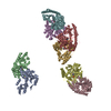
| ||||||||
|---|---|---|---|---|---|---|---|---|---|
| 1 | 
| ||||||||
| 2 | 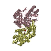
| ||||||||
| 3 | 
| ||||||||
| 4 | 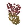
| ||||||||
| Unit cell |
|
-Components
| #1: Protein | Mass: 46278.441 Da / Num. of mol.: 8 Source method: isolated from a genetically manipulated source Source: (gene. exp.) Treponema denticola (bacteria) / Plasmid: PLC67 / Production host: #2: Chemical | ChemComp-PLP /   Mass: 247.142 Da / Num. of mol.: 8 / Source method: obtained synthetically / Formula: C8H10NO6P Mass: 247.142 Da / Num. of mol.: 8 / Source method: obtained synthetically / Formula: C8H10NO6P#3: Water | ChemComp-HOH / |  Mass: 18.015 Da / Num. of mol.: 2626 / Source method: isolated from a natural source / Formula: H2O Mass: 18.015 Da / Num. of mol.: 2626 / Source method: isolated from a natural source / Formula: H2O |
|---|
-Experimental details
-Experiment
| Experiment | Method: X-RAY DIFFRACTION / Number of used crystals: 1 |
|---|
- Sample preparation
| Crystal | Density Matthews: 2.31 Å3/Da / Density % sol: 46.67 % | |||||||||||||||||||||||||
|---|---|---|---|---|---|---|---|---|---|---|---|---|---|---|---|---|---|---|---|---|---|---|---|---|---|---|
| Crystal grow | Temperature: 293 K / Method: vapor diffusion, sitting drop / pH: 6.5 Details: PEG 4000, magnesium acetate, MES, pH 6.5, vapor diffusion/sitting drop, temperature 293.0K | |||||||||||||||||||||||||
| Crystal grow | *PLUS Temperature: 20 ℃ / Method: vapor diffusion, sitting drop | |||||||||||||||||||||||||
| Components of the solutions | *PLUS
|
-Data collection
| Diffraction | Mean temperature: 100 K |
|---|---|
| Diffraction source | Source: SYNCHROTRON / Site: MPG/DESY, HAMBURG  / Beamline: BW6 / Wavelength: 1.07 / Beamline: BW6 / Wavelength: 1.07 |
| Detector | Type: MARRESEARCH / Detector: CCD / Date: Nov 8, 1998 |
| Radiation | Protocol: SINGLE WAVELENGTH / Monochromatic (M) / Laue (L): M / Scattering type: x-ray |
| Radiation wavelength | Wavelength: 1.07 Å / Relative weight: 1 |
| Reflection | Resolution: 1.9→25 Å / Num. all: 382434 / Num. obs: 254989 / % possible obs: 96.5 % / Redundancy: 1.5 % / Biso Wilson estimate: 16.9 Å2 / Rmerge(I) obs: 0.078 / Net I/σ(I): 8.6 |
| Reflection shell | Resolution: 1.9→1.97 Å / Redundancy: 1.6 % / Rmerge(I) obs: 0.255 / Num. unique all: 12280 / % possible all: 87.9 |
| Reflection | *PLUS Num. measured all: 382434 |
| Reflection shell | *PLUS % possible obs: 87.9 % |
- Processing
| Refinement | Resolution: 1.9→25 Å / Stereochemistry target values: Engh & Huber
| |||||||||||||||||||||||||
|---|---|---|---|---|---|---|---|---|---|---|---|---|---|---|---|---|---|---|---|---|---|---|---|---|---|---|
| Refinement step | Cycle: LAST / Resolution: 1.9→25 Å
| |||||||||||||||||||||||||
| Refinement | *PLUS | |||||||||||||||||||||||||
| Solvent computation | *PLUS | |||||||||||||||||||||||||
| Displacement parameters | *PLUS Biso mean: 16.9 Å2 | |||||||||||||||||||||||||
| Refine LS restraints | *PLUS
|
