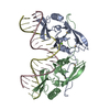
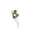
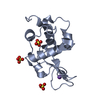
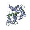
PDB-9gfu: Human PPAR-gamma ligand binding domain in complex with AKGO172 Method: X-RAY DIFFRACTION / Resolution: 2.1 Å
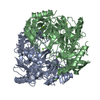
PDB-9gvp: Human PPAR-gamma ligand binding domain in complex with WG115 Method: X-RAY DIFFRACTION / Resolution: 2.05 Å
PDB-9gvs: Human PPAR-gamma ligand binding domain in complex with AK176_C Method: X-RAY DIFFRACTION / Resolution: 2.1 Å
PDB-9gw3: Human PPAR-gamma ligand binding domain in complex with LW56 Method: X-RAY DIFFRACTION / Resolution: 2 Å
PDB-9gw4: Human PPAR-gamma ligand binding domain in complex with LW78 Method: X-RAY DIFFRACTION / Resolution: 2.2 Å
PDB-9gw7: Human PPAR-gamma ligand binding domain in complex with LW76 Method: X-RAY DIFFRACTION / Resolution: 2.1 Å
PDB-9gw8: Human PPAR-gamma ligand binding domain in complex with LW75 Method: X-RAY DIFFRACTION / Resolution: 2.2 Å
PDB-9gwb: Human PPAR-gamma ligand binding domain in complex with LW69 Method: X-RAY DIFFRACTION / Resolution: 2.2 Å
PDB-9gwc: Human PPAR-gamma ligand binding domain in complex with LW99 Method: X-RAY DIFFRACTION / Resolution: 2 Å
PDB-9gwe: Human PPAR-gamma ligand binding domain in complex with LW87 Method: X-RAY DIFFRACTION / Resolution: 2.1 Å
PDB-9gwf: Human PPAR-gamma ligand binding domain in complex with LW100 Method: X-RAY DIFFRACTION / Resolution: 2.4 Å
PDB-9gwg: Human PPAR-gamma ligand binding domain in complex with LW88 Method: X-RAY DIFFRACTION / Resolution: 2.3 Å
PDB-9gwh: Human PPAR-gamma ligand binding domain in complex with LW77 Method: X-RAY DIFFRACTION / Resolution: 2.1 Å
PDB-9gwi: Human PPAR-gamma ligand binding domain in complex with LW23 Method: X-RAY DIFFRACTION / Resolution: 2.2 Å
PDB-9gwk: Human PPAR-gamma ligand binding domain in complex with AKGO6A Method: X-RAY DIFFRACTION / Resolution: 2.2 Å
PDB-9gwl: Human PPAR-gamma ligand binding domain in complex with AKGO157 Method: X-RAY DIFFRACTION / Resolution: 2.2 Å
PDB-9gwm: Human PPAR-gamma ligand binding domain in complex with AKGO108 Method: X-RAY DIFFRACTION / Resolution: 2.2 Å
PDB-9gwn: Human PPAR-gamma ligand binding domain in complex with LW30 Method: X-RAY DIFFRACTION / Resolution: 2.2 Å
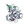
PDB-9gwp: Human PPAR-gamma ligand binding domain in complex with MRL-871 with two distinct binding modes Method: X-RAY DIFFRACTION / Resolution: 2.55 Å
PDB-9gwq: Human PPAR-gamma ligand binding domain in complex with AKGO6D Method: X-RAY DIFFRACTION / Resolution: 2.1 Å
PDB-9gwr: Human PPAR-gamma ligand binding domain in complex with AKGO10 Method: X-RAY DIFFRACTION / Resolution: 2.1 Å
PDB-9gws: Human PPAR-gamma ligand binding domain in complex with AKGO8 Method: X-RAY DIFFRACTION / Resolution: 2.2 Å
PDB-9gwx: Human PPAR-gamma ligand binding domain in complex with AKGO141 Method: X-RAY DIFFRACTION / Resolution: 2.2 Å
PDB-9gwy: Human PPAR-gamma ligand binding domain in complex with AKGO78 Method: X-RAY DIFFRACTION / Resolution: 2.09 Å
PDB-9gx0: Human PPAR-gamma ligand binding domain in complex with AKGO79 Method: X-RAY DIFFRACTION / Resolution: 2.1 Å
PDB-9gx1: Human PPAR-gamma ligand binding domain in complex with AKGO80 Method: X-RAY DIFFRACTION / Resolution: 2.1 Å
PDB-9gx2: Human PPAR-gamma ligand binding domain in complex with AKGO82 Method: X-RAY DIFFRACTION / Resolution: 2.35 Å
PDB-9gx3: Human PPAR-gamma ligand binding domain in complex with AKGO12 Method: X-RAY DIFFRACTION / Resolution: 1.9 Å
PDB-9gx4: Human PPAR-gamma ligand binding domain in complex with AKGO156 Method: X-RAY DIFFRACTION / Resolution: 2.2 Å
PDB-9gx5: Human PPAR-gamma ligand binding domain in complex with AKGO173 Method: X-RAY DIFFRACTION / Resolution: 2.45 Å
PDB-9gx6: Human PPAR-gamma ligand binding domain in complex with AKGO166 Method: X-RAY DIFFRACTION / Resolution: 2.1 Å
PDB-9gx7: Human PPAR-gamma ligand binding domain in complex with WG17 Method: X-RAY DIFFRACTION / Resolution: 2.2 Å
PDB-9gxd: Human PPAR-gamma ligand binding domain in complex with AKGO27 Method: X-RAY DIFFRACTION / Resolution: 2.2 Å
PDB-9gz7: Human PPAR-gamma ligand binding domain in complex with AKGO29 Method: X-RAY DIFFRACTION / Resolution: 1.8 Å
PDB-9gz8: Human PPAR-gamma ligand binding domain in complex with AKGO46 Method: X-RAY DIFFRACTION / Resolution: 2.2 Å
PDB-9gz9: Human PPAR-gamma ligand binding domain in complex with AKGO48 Method: X-RAY DIFFRACTION / Resolution: 2.1 Å
PDB-9gza: Human PPAR-gamma ligand binding domain in complex with AKGO50 Method: X-RAY DIFFRACTION / Resolution: 1.9 Å
PDB-9gzb: Human PPAR-gamma ligand binding domain in complex with AKGO52 Method: X-RAY DIFFRACTION / Resolution: 2.2 Å
PDB-9gzc: Human PPAR-gamma ligand binding domain in complex with AKGO142 Method: X-RAY DIFFRACTION / Resolution: 2.3 Å
PDB-9gze: Human PPAR-gamma ligand binding domain in complex with AKGO72 Method: X-RAY DIFFRACTION / Resolution: 2.2 Å
PDB-9gzf: Human PPAR-gamma ligand binding domain in complex with AKGO88 Method: X-RAY DIFFRACTION / Resolution: 2.65 Å
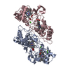
PDB-9gzi: Human PPAR-gamma ligand binding domain in complex with LW98 Method: X-RAY DIFFRACTION / Resolution: 2.7 Å
Chemicals
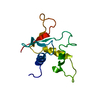
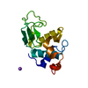
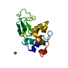
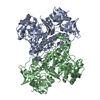
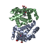
PDB-1ik6: 3D structure of the E1beta subunit of pyruvate dehydrogenase from the archeon Pyrobaculum aerophilum
In the structure databanks used in Yorodumi, some data are registered as the other names, "COVID-19 virus" and "2019-nCoV". Here are the details of the virus and the list of structure data.
Jan 31, 2019. EMDB accession codes are about to change! (news from PDBe EMDB page)
EMDB accession codes are about to change! (news from PDBe EMDB page)
The allocation of 4 digits for EMDB accession codes will soon come to an end. Whilst these codes will remain in use, new EMDB accession codes will include an additional digit and will expand incrementally as the available range of codes is exhausted. The current 4-digit format prefixed with “EMD-” (i.e. EMD-XXXX) will advance to a 5-digit format (i.e. EMD-XXXXX), and so on. It is currently estimated that the 4-digit codes will be depleted around Spring 2019, at which point the 5-digit format will come into force.
The EM Navigator/Yorodumi systems omit the EMD- prefix.
Related info.:Q: What is EMD? / ID/Accession-code notation in Yorodumi/EM Navigator
 Movie
Movie Controller
 Structure viewers
 About Yorodumi Papers


 Authors
 External links


 Keywords
 homo sapiens (human)