 Movie
Movie Controller


+ Open data

- Basic information
| Entry | Database: PDB / ID: 1iq6 | ||||||
|---|---|---|---|---|---|---|---|
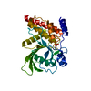
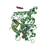
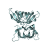
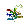
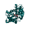
| Title | (R)-HYDRATASE FROM A. CAVIAE INVOLVED IN PHA BIOSYNTHESIS | ||||||
 Components Components | (R)-SPECIFIC ENOYL-COA HYDRATASE | ||||||
 Keywords Keywords | LYASE / hydratase / (R)-hydratase / enoyl-CoA hydratase / polyhydroxyalkanoate / Aeromonas caviae / the hydratase 2 motif | ||||||
| Function / homology |  Function and homology information Function and homology informationenoyl-CoA hydratase 2 / 3-hydroxyacyl-CoA dehydratase activity / fatty acid synthase complex / (3R)-hydroxyacyl-[acyl-carrier-protein] dehydratase activity / fatty acid synthase activity / fatty acid biosynthetic process Similarity search - Function | ||||||
| Biological species |  Aeromonas punctata (bacteria) Aeromonas punctata (bacteria) | ||||||
| Method |  X-RAY DIFFRACTION / SYNCHROTRON / MIRAS / Resolution: 1.5 Å X-RAY DIFFRACTION / SYNCHROTRON / MIRAS / Resolution: 1.5 Å | ||||||
 Authors Authors | Hisano, T. / Tsuge, T. / Fukui, T. / Iwata, T. / Doi, Y. | ||||||
 Citation Citation | Journal: J.Biol.Chem. / Year: 2003 Title: Crystal structure of the (R)-specific enoyl-CoA hydratase from Aeromonas caviae involved in polyhydroxyalkanoate biosynthesis Authors: Hisano, T. / Tsuge, T. / Fukui, T. / Iwata, T. / Miki, K. / Doi, Y. #1: Journal: Acta Crystallogr.,Sect.D / Year: 2001Title: CRYSTALLIZATION AND PRELIMINARY X-RAY ANALYSIS OF (R)-SPECIFIC ENOYL-COA HYDRATASE FROM AEROMONAS CAVIAE INVOLVED IN POLYHYDROXYALKANOATE BIOSYNTHESIS Authors: Hisano, T. / Fukui, T. / Iwata, T. / Doi, Y. #2: Journal: J.BACTERIOL. / Year: 1998Title: EXPRESSION AND CHARACTERIZATION OF (R)-SPECIFIC ENOYL-COA HYDRATASE INVOLVED IN POLYHYDROXYALKANOATE BIOSYNTHESIS BY AEROMONAS CAVIAE Authors: Fukui, T. / SHIOMI, N. / Doi, Y. #3: Journal: J.BACTERIOL. / Year: 1997Title: CLONING AND ANALYSIS OF THE POLY(3-HYDROXYBUTYRATE-CO-3-HYDROXYHEXANOATE) BIOSYNTHESIS GENES OF AEROMONAS CAVIAE Authors: Fukui, T. / Doi, Y. | ||||||
| History |
|
- Structure visualization
| Structure viewer | Molecule: MolmilJmol/JSmol |
|---|
- Downloads & links
-Download
| PDBx/mmCIF format | 1iq6.cif.gz | 68.3 KB | Display | PDBx/mmCIF format |
|---|---|---|---|---|
| PDB format | pdb1iq6.ent.gz | 50.7 KB | Display | PDB format |
| PDBx/mmJSON format | 1iq6.json.gz | Tree view | PDBx/mmJSON format | |
| Others |  Other downloads Other downloads |
-Validation report
| Arichive directory | https://data.pdbj.org/pub/pdb/validation_reports/iq/1iq6ftp://data.pdbj.org/pub/pdb/validation_reports/iq/1iq6 | HTTPS FTP |
|---|
-Related structure data
| Similar structure data |
|---|

-Links
 PDBj
PDBj

- Assembly
| Deposited unit | 
| ||||||||
|---|---|---|---|---|---|---|---|---|---|
| 1 |
| ||||||||
| Unit cell |
|
-Components
| #1: Protein | Mass: 14099.186 Da / Num. of mol.: 2 Source method: isolated from a genetically manipulated source Source: (gene. exp.) Aeromonas punctata (bacteria) / Plasmid: PET3A / Species (production host): Escherichia coli / Production host: #2: Chemical | ChemComp-IPA / |   Mass: 60.095 Da / Num. of mol.: 1 / Source method: obtained synthetically / Formula: C3H8O / Comment: alkaloid*YM Mass: 60.095 Da / Num. of mol.: 1 / Source method: obtained synthetically / Formula: C3H8O / Comment: alkaloid*YM#3: Water | ChemComp-HOH / |  Mass: 18.015 Da / Num. of mol.: 347 / Source method: isolated from a natural source / Formula: H2O Mass: 18.015 Da / Num. of mol.: 347 / Source method: isolated from a natural source / Formula: H2O |
|---|
-Experimental details
-Experiment
| Experiment | Method: X-RAY DIFFRACTION / Number of used crystals: 1 |
|---|
- Sample preparation
| Crystal | Density Matthews: 2.44 Å3/Da / Density % sol: 49.63 % Description: THE INITIAL STRUCTURE WAS DETERMINED AT 3.0 A RESOLUTION BY MIRAS WITH NATIVE DATA AND THREE SETS OF DERIVATIVE DATA, ALL OF WHICH WERE COLLECTED AT ROOM TEMPERATURE WITH A ROTATING ...Description: THE INITIAL STRUCTURE WAS DETERMINED AT 3.0 A RESOLUTION BY MIRAS WITH NATIVE DATA AND THREE SETS OF DERIVATIVE DATA, ALL OF WHICH WERE COLLECTED AT ROOM TEMPERATURE WITH A ROTATING ANODE X-RAY GENERATOR. THIS MODEL WAS USED TO PHASE THE 1.5 A DATA COLLECTED AT 100K WITH SYNCHROTRON RADIATION BY MOLECULAR REPLACEMENT, AND REFINED BY MOLECULAR DYNAMICS SIMULATION. | ||||||||||||||||||||||||
|---|---|---|---|---|---|---|---|---|---|---|---|---|---|---|---|---|---|---|---|---|---|---|---|---|---|
| Crystal grow | Temperature: 298 K / Method: vapor diffusion, sitting drop / pH: 6 Details: PEG4000, HEPES, ISOPROPANOL, pH 6.00, VAPOR DIFFUSION, SITTING DROP, temperature 298K | ||||||||||||||||||||||||
| Crystal grow | *PLUS Temperature: 25 ℃ / pH: 7 | ||||||||||||||||||||||||
| Components of the solutions | *PLUS
|
-Data collection
| Diffraction | Mean temperature: 100 K |
|---|---|
| Diffraction source | Source: SYNCHROTRON / Site: SPring-8  / Beamline: BL44B2 / Wavelength: 1 Å / Beamline: BL44B2 / Wavelength: 1 Å |
| Detector | Type: MARRESEARCH / Detector: CCD |
| Radiation | Protocol: SINGLE WAVELENGTH / Monochromatic (M) / Laue (L): M / Scattering type: x-ray |
| Radiation wavelength | Wavelength: 1 Å / Relative weight: 1 |
| Reflection | Highest resolution: 2.5 Å / Num. obs: 52985 / % possible obs: 99.4 % / Biso Wilson estimate: 20.2 Å2 / Rmerge(I) obs: 0.052 |
| Reflection | *PLUS Highest resolution: 1.5 Å / Num. obs: 52985 / % possible obs: 99.4 % / Num. measured all: 198695 / Rmerge(I) obs: 0.052 |
- Processing
| Software |
| ||||||||||||||||||||||||||||||||||||||||||||||||||||||||||||||||||||||||||||||||
|---|---|---|---|---|---|---|---|---|---|---|---|---|---|---|---|---|---|---|---|---|---|---|---|---|---|---|---|---|---|---|---|---|---|---|---|---|---|---|---|---|---|---|---|---|---|---|---|---|---|---|---|---|---|---|---|---|---|---|---|---|---|---|---|---|---|---|---|---|---|---|---|---|---|---|---|---|---|---|---|---|---|
| Refinement | Method to determine structure: MIRAS Starting model: A STRUCTURE OF (R)-HYDRATASE FROM A.CAVIAE DETERMINED BY MIRAS AT 3.0 A RESOLUTION Resolution: 1.5→19.15 Å / Rfactor Rfree error: 0.005 / Cross valid method: THROUGHOUT / σ(F): 0
| ||||||||||||||||||||||||||||||||||||||||||||||||||||||||||||||||||||||||||||||||
| Solvent computation | Solvent model: FLAT MODEL / Bsol: 62.65 Å2 / ksol: 0.43 e/Å3 | ||||||||||||||||||||||||||||||||||||||||||||||||||||||||||||||||||||||||||||||||
| Displacement parameters | Biso mean: 21.4 Å2
| ||||||||||||||||||||||||||||||||||||||||||||||||||||||||||||||||||||||||||||||||
| Refine analyze |
| ||||||||||||||||||||||||||||||||||||||||||||||||||||||||||||||||||||||||||||||||
| Refinement step | Cycle: LAST / Resolution: 1.5→19.15 Å
| ||||||||||||||||||||||||||||||||||||||||||||||||||||||||||||||||||||||||||||||||
| Refine LS restraints |
| ||||||||||||||||||||||||||||||||||||||||||||||||||||||||||||||||||||||||||||||||
| LS refinement shell | Resolution: 1.5→1.59 Å / Rfactor Rfree error: 0.013 / Total num. of bins used: 6
| ||||||||||||||||||||||||||||||||||||||||||||||||||||||||||||||||||||||||||||||||
| Refinement | *PLUS Highest resolution: 1.5 Å / Lowest resolution: 19.15 Å / % reflection Rfree: 5 % | ||||||||||||||||||||||||||||||||||||||||||||||||||||||||||||||||||||||||||||||||
| Solvent computation | *PLUS | ||||||||||||||||||||||||||||||||||||||||||||||||||||||||||||||||||||||||||||||||
| Displacement parameters | *PLUS | ||||||||||||||||||||||||||||||||||||||||||||||||||||||||||||||||||||||||||||||||
| Refine LS restraints | *PLUS
|
