- PDB-3irb: Crystal structure of protein with unknown function from DUF35 fam... -
+
Open data
ID or keywords:
Loading...
-
Basic information
Entry
Database: PDB / ID: 3irb
Title
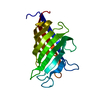
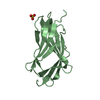
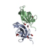
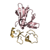
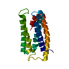
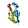
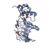
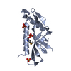
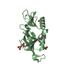
Crystal structure of protein with unknown function from DUF35 family (13815350) from SULFOLOBUS SOLFATARICUS at 1.80 A resolution
Components
uncharacterized protein from DUF35 family
Keywords
acyl-CoA binding protein / 13815350 / protein with unknown function from DUF35 family / Structural Genomics / Joint Center for Structural Genomics / JCSG / Protein Structure Initiative / PSI-2 / Domain of unknown function DUF35 / unknown function
SIZE EXCLUSION CHROMATOGRAPHY WITH STATIC LIGHT SCATTERING SUPPORTS THE ASSIGNMENT OF A MONOMER AS A SIGNIFICANT OLIGOMERIZATION STATE IN SOLUTION. CRYSTAL PACKING SUGGESTS A POSSIBLE DIMER OR TETRAMER IN THE CRYSTAL.
-
Components
#1: Protein
uncharacterizedproteinfromDUF35family
Mass: 16637.693 Da / Num. of mol.: 2 Source method: isolated from a genetically manipulated source Source: (gene. exp.) Sulfolobus solfataricus (archaea) / Gene: 13815350, SSO2064 / Plasmid: SpeedET / Production host: Escherichia Coli (E. coli) / Strain (production host): HK100 / References: UniProt: Q97WQ4
Mass: 18.015 Da / Num. of mol.: 201 / Source method: isolated from a natural source / Formula: H2O
Has protein modification
Y
Sequence details
SEQUENCE THE CONSTRUCT WAS EXPRESSED WITH A PURIFICATION TAG MGSDKIHHHHHHENLYFQ/G. THE TAG WAS ...SEQUENCE THE CONSTRUCT WAS EXPRESSED WITH A PURIFICATION TAG MGSDKIHHHHHHENLYFQ/G. THE TAG WAS REMOVED WITH TEV PROTEASE LEAVING ONLY A GLYCINE (0) FOLLOWED BY THE TARGET SEQUENCE.
-
Experimental details
-
Experiment
Experiment
Method: X-RAY DIFFRACTION / Number of used crystals: 1
-
Sample preparation
Crystal
Density Matthews: 2.45 Å3/Da / Density % sol: 49.79 %
Crystal grow
Temperature: 277 K / Method: vapor diffusion, sitting drop / pH: 4.6 Details: 2.0M (NH4)2SO4, 0.1M Acetate pH 4.6, NANODROP, VAPOR DIFFUSION, SITTING DROP, temperature 277K
Monochromator: Single crystal Si(111) bent monochromator (horizontal focusing) Protocol: MAD / Monochromatic (M) / Laue (L): M / Scattering type: x-ray
Radiation wavelength
ID
Wavelength (Å)
Relative weight
1
0.91837
1
2
0.979386
1
3
0.97917
1
Reflection
Resolution: 1.8→26.939 Å / Num. obs: 31362 / % possible obs: 99.2 % / Observed criterion σ(I): -3 / Redundancy: 7.056 % / Biso Wilson estimate: 24.984 Å2 / Rmerge(I) obs: 0.081 / Net I/σ(I): 9.73
Reflection shell
Resolution (Å)
Rmerge(I) obs
Mean I/σ(I) obs
Num. measured obs
Num. unique obs
Diffraction-ID
% possible all
1.8-1.86
0.792
1.9
19058
5114
1
93
1.86-1.94
0.618
2.5
24071
6369
1
99.9
1.94-2.03
0.445
3.4
22540
5952
1
99.9
2.03-2.13
0.32
4.5
20951
5521
1
99.9
2.13-2.27
0.223
6.3
23487
6182
1
100
2.27-2.44
0.184
7.5
21646
5690
1
99.9
2.44-2.69
0.145
9
22823
5995
1
99.9
2.69-3.07
0.098
12
21968
5776
1
99.9
3.07-3.87
0.056
19.2
22462
5938
1
99.9
3.87-26.939
0.031
30.1
22287
5928
1
99.3
-
Phasing
Phasing
Method: MAD
-
Processing
Software
Name
Version
Classification
NB
REFMAC
5.5.0099
refinement
PHENIX
refinement
SHELX
phasing
MolProbity
3beta29
modelbuilding
XSCALE
datascaling
PDB_EXTRACT
3.006
dataextraction
XDS
datareduction
SHELXD
phasing
autoSHARP
phasing
Refinement
Method to determine structure: MAD / Resolution: 1.8→26.939 Å / Cor.coef. Fo:Fc: 0.965 / Cor.coef. Fo:Fc free: 0.953 / Occupancy max: 1 / Occupancy min: 0.35 / SU B: 5.468 / SU ML: 0.075 / TLS residual ADP flag: LIKELY RESIDUAL / Cross valid method: THROUGHOUT / σ(F): 0 / ESU R: 0.108 / ESU R Free: 0.105 Stereochemistry target values: MAXIMUM LIKELIHOOD WITH PHASES Details: 1. HYDROGENS HAVE BEEN ADDED IN THE RIDING POSITIONS. 2. ATOM RECORD CONTAINS RESIDUAL B FACTORS ONLY. 3. A MET-INHIBITION PROTOCOL WAS USED FOR SELENOMETHIONINE INCORPORATION DURING PROTEIN ...Details: 1. HYDROGENS HAVE BEEN ADDED IN THE RIDING POSITIONS. 2. ATOM RECORD CONTAINS RESIDUAL B FACTORS ONLY. 3. A MET-INHIBITION PROTOCOL WAS USED FOR SELENOMETHIONINE INCORPORATION DURING PROTEIN EXPRESSION. THE OCCUPANCY OF THE SE ATOMS IN THE MSE RESIDUES WAS REDUCED TO 0.75 FOR THE REDUCED SCATTERING POWER DUE TO PARTIAL S-MET INCORPORATION. 4. ZINC IONS ARE MODELED IN THE CONSERVED RUBREDOXIN DOMAIN ZINC BINDING SITE IN EACH CHAIN. THE PRESENCE OF ZINC IS SUPPORTED BY X-RAY FLUORESCENCE EXCITATION AND WAVELENGTH SCANS, ANOMALOUS DIFFERENCE FOURIERS AND COORDINATION GEOMETRY. 5. ACETATE (ACY) AND SULFATE IONS (SO4) FROM THE CRYSTALLIZATION CONDITIONS ARE MODELED.
Rfactor
Num. reflection
% reflection
Selection details
Rfree
0.2
1560
5 %
RANDOM
Rwork
0.169
-
-
-
obs
0.171
31317
99.77 %
-
Solvent computation
Ion probe radii: 0.8 Å / Shrinkage radii: 0.8 Å / VDW probe radii: 1.4 Å / Solvent model: MASK
In the structure databanks used in Yorodumi, some data are registered as the other names, "COVID-19 virus" and "2019-nCoV". Here are the details of the virus and the list of structure data.
Jan 31, 2019. EMDB accession codes are about to change! (news from PDBe EMDB page)
EMDB accession codes are about to change! (news from PDBe EMDB page)
The allocation of 4 digits for EMDB accession codes will soon come to an end. Whilst these codes will remain in use, new EMDB accession codes will include an additional digit and will expand incrementally as the available range of codes is exhausted. The current 4-digit format prefixed with “EMD-” (i.e. EMD-XXXX) will advance to a 5-digit format (i.e. EMD-XXXXX), and so on. It is currently estimated that the 4-digit codes will be depleted around Spring 2019, at which point the 5-digit format will come into force.
The EM Navigator/Yorodumi systems omit the EMD- prefix.
Related info.:Q: What is EMD? / ID/Accession-code notation in Yorodumi/EM Navigator
Yorodumi is a browser for structure data from EMDB, PDB, SASBDB, etc.
This page is also the successor to EM Navigator detail page, and also detail information page/front-end page for Omokage search.
The word "yorodu" (or yorozu) is an old Japanese word meaning "ten thousand". "mi" (miru) is to see.
Related info.:EMDB / PDB / SASBDB / Comparison of 3 databanks / Yorodumi Search / Aug 31, 2016. New EM Navigator & Yorodumi / Yorodumi Papers / Jmol/JSmol / Function and homology information / Changes in new EM Navigator and Yorodumi
 Movie
Movie Controller

 Yorodumi
 Open data
 Basic information
 Components
 Keywords
 Function and homology information
 Sulfolobus solfataricus (archaea)
 X-RAY DIFFRACTION /
  Authors
 Citation
 Structure visualization
 Downloads & links
 Other downloads
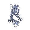


 PDBj
PDBj

 Assembly
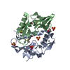


 Mass: 65.409 Da / Num. of mol.: 2 / Source method: obtained synthetically / Formula: Zn
 Mass: 96.063 Da / Num. of mol.: 6 / Source method: obtained synthetically / Formula: SO4
 Mass: 60.052 Da / Num. of mol.: 2 / Source method: obtained synthetically / Formula: C2H4O2
 Mass: 18.015 Da / Num. of mol.: 201 / Source method: isolated from a natural source / Formula: H2O
 Sample preparation
 / Beamline: BL11-1 / Wavelength: 0.918370,0.979386,0.979170
 Processing