Mass: 18.015 Da / Num. of mol.: 229 / Source method: isolated from a natural source / Formula: H2O
Sequence details
THE M231V DIFFERENCE IS OWING TO STRAIN VARIATION
-
Experimental details
-
Experiment
Experiment
Method: X-RAY DIFFRACTION / Number of used crystals: 1
-
Sample preparation
Crystal
Density Matthews: 2.1 Å3/Da / Density % sol: 41.45 % / Description: NONE
Crystal grow
Details: 0.2 M POTASSIUM FORMATE, 30% PEG 3350 AND A PROTEIN SOLUTION CONSISTING OF 50 MM TRIS-HCL PH 7.5, 250 MM NABR WITH THE PROTEIN AT 7.5 MG ML-1.
Resolution: 1.59→45.79 Å / Cor.coef. Fo:Fc: 0.946 / Cor.coef. Fo:Fc free: 0.921 / SU B: 1.93 / SU ML: 0.07 / Cross valid method: THROUGHOUT / ESU R: 0.112 / ESU R Free: 0.11 / Stereochemistry target values: MAXIMUM LIKELIHOOD Details: HYDROGENS HAVE BEEN ADDED IN THE RIDING POSITIONS. RESIDUES 149 TO 152 AND TWO RESIDUES LEFT OVER FROM THE TAG DISORDERED IN BOTH CHAINS. FINAL RESIDUE 232 OF CHAIN B DISORDERED
Rfactor
Num. reflection
% reflection
Selection details
Rfree
0.24034
1042
5.1 %
RANDOM
Rwork
0.20094
-
-
-
obs
0.20286
19291
99.96 %
-
Solvent computation
Ion probe radii: 0.8 Å / Shrinkage radii: 0.8 Å / VDW probe radii: 1.2 Å / Solvent model: MASK
 Movie
Movie Controller


 Open data
 Basic information
 Components
 Keywords
 Function and homology information
 X-RAY DIFFRACTION /
  Authors
 Citation
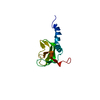
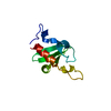
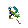
 Structure visualization
 Downloads & links
 Other downloads
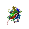


 PDBj
PDBj Assembly


 Mass: 46.025 Da / Num. of mol.: 7 / Source method: obtained synthetically / Formula: CH2O2
 Mass: 18.015 Da / Num. of mol.: 229 / Source method: isolated from a natural source / Formula: H2O
 Sample preparation
 / Beamline: I02 / Wavelength: 0.9199
  Processing