 Movie
Movie Controller

[English] 日本語
 Yorodumi
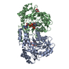
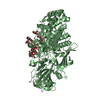
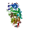
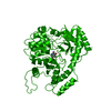
- PDB-1iqc: Crystal structure of Di-Heme Peroxidase from Nitrosomonas europaea -
+ Open data

- Basic information
| Entry | Database: PDB / ID: 1iqc | |||||||||
|---|---|---|---|---|---|---|---|---|---|---|
| Title | Crystal structure of Di-Heme Peroxidase from Nitrosomonas europaea | |||||||||
 Components Components | di-heme peroxidase | |||||||||
 Keywords Keywords | OXIDOREDUCTASE / Di-Heme Peroxidase / Nitrosomonas europaea / Proteobacteria / beta subdivision / Ammonia-oxidizing bacteria | |||||||||
| Function / homology |  Function and homology information Function and homology informationcytochrome-c peroxidase / cytochrome-c peroxidase activity / electron transfer activity / periplasmic space / heme binding / metal ion binding Similarity search - Function | |||||||||
| Biological species |  Nitrosomonas europaea (bacteria) Nitrosomonas europaea (bacteria) | |||||||||
| Method |  X-RAY DIFFRACTION / SYNCHROTRON / MOLECULAR REPLACEMENT / Resolution: 1.8 Å X-RAY DIFFRACTION / SYNCHROTRON / MOLECULAR REPLACEMENT / Resolution: 1.8 Å | |||||||||
 Authors Authors | Shimizu, H. | |||||||||
 Citation Citation | Journal: Biochemistry / Year: 2001 Title: Crystal structure of Nitrosomonas europaea cytochrome c peroxidase and the structural basis for ligand switching in bacterial di-heme peroxidases Authors: Shimizu, H. / Schuller, D.J. / Lanzilotta, W.N. / Sundaramoorthy, M. / Arciero, D.M. / Hooper, A.B. / Poulos, T.L. #1: Journal: J.STRUCT.BIOL. / Year: 1996Title: Crystallization and Preliminary Crystallographic Analysis of Cytochrome C553 Peroxidase from Nitrosomonas europaea Authors: Pappa, H. / Li, H. / Sundaramoorthy, M. / Arciero, D. / Hooper, A. / Poulos, T.L. | |||||||||
| History |
|
- Structure visualization
| Structure viewer | Molecule: MolmilJmol/JSmol |
|---|
- Downloads & links
-Download
| PDBx/mmCIF format | 1iqc.cif.gz | 276.4 KB | Display | PDBx/mmCIF format |
|---|---|---|---|---|
| PDB format | pdb1iqc.ent.gz | 222.9 KB | Display | PDB format |
| PDBx/mmJSON format | 1iqc.json.gz | Tree view | PDBx/mmJSON format | |
| Others |  Other downloads Other downloads |
-Validation report
| Arichive directory | https://data.pdbj.org/pub/pdb/validation_reports/iq/1iqcftp://data.pdbj.org/pub/pdb/validation_reports/iq/1iqc | HTTPS FTP |
|---|
-Related structure data
| Similar structure data |
|---|


-Links
 PDBj
PDBj


- Assembly
| Deposited unit | 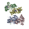
| ||||||||
|---|---|---|---|---|---|---|---|---|---|
| 1 | 
| ||||||||
| 2 | 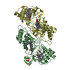
| ||||||||
| Unit cell |
|
-Components
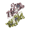
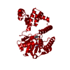
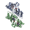
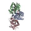
-Protein , 1 types, 4 molecules ABCD
| #1: Protein | Mass: 34019.191 Da / Num. of mol.: 4 / Source method: isolated from a natural source / Source: (natural) Nitrosomonas europaea (bacteria) / References: UniProt: P55929 |
|---|
-Non-polymers , 5 types, 874 molecules 


| #2: Chemical | ChemComp-CA /  Mass: 40.078 Da / Num. of mol.: 5 / Source method: obtained synthetically / Formula: Ca Mass: 40.078 Da / Num. of mol.: 5 / Source method: obtained synthetically / Formula: Ca#3: Chemical |  Mass: 24.305 Da / Num. of mol.: 2 / Source method: obtained synthetically / Formula: Mg Mass: 24.305 Da / Num. of mol.: 2 / Source method: obtained synthetically / Formula: Mg#4: Chemical | ChemComp-HEC /  Mass: 618.503 Da / Num. of mol.: 8 / Source method: obtained synthetically / Formula: C34H34FeN4O4 Mass: 618.503 Da / Num. of mol.: 8 / Source method: obtained synthetically / Formula: C34H34FeN4O4#5: Chemical | ChemComp-GOL / |  Mass: 92.094 Da / Num. of mol.: 1 / Source method: obtained synthetically / Formula: C3H8O3 Mass: 92.094 Da / Num. of mol.: 1 / Source method: obtained synthetically / Formula: C3H8O3#6: Water | ChemComp-HOH / | Mass: 18.015 Da / Num. of mol.: 858 / Source method: isolated from a natural source / Formula: H2O |
|---|
-Details
| Has protein modification | Y |
|---|
-Experimental details
-Experiment
| Experiment | Method: X-RAY DIFFRACTION / Number of used crystals: 1 |
|---|
- Sample preparation
| Crystal | Density Matthews: 2.5 Å3/Da / Density % sol: 50.73 % | |||||||||||||||||||||||||||||||||||
|---|---|---|---|---|---|---|---|---|---|---|---|---|---|---|---|---|---|---|---|---|---|---|---|---|---|---|---|---|---|---|---|---|---|---|---|---|
| Crystal grow | Temperature: 298 K / Method: vapor diffusion, hanging drop / pH: 8.5 Details: PEG8000, MgCl2, Tris-HCl, pH 8.5, VAPOR DIFFUSION, HANGING DROP, temperature 298K | |||||||||||||||||||||||||||||||||||
| Crystal grow | *PLUS Details: Pappa, H., (1996) J.STRUCT.BIOL., 116, 429. | |||||||||||||||||||||||||||||||||||
| Components of the solutions | *PLUS
|
-Data collection
| Diffraction | Mean temperature: 100 K |
|---|---|
| Diffraction source | Source: SYNCHROTRON / Site: ALS  / Beamline: 5.0.2 / Wavelength: 1 Å / Beamline: 5.0.2 / Wavelength: 1 Å |
| Detector | Type: ADSC QUANTUM 4 / Detector: CCD / Date: Jun 15, 2000 |
| Radiation | Protocol: MAD / Monochromatic (M) / Laue (L): M / Scattering type: x-ray |
| Radiation wavelength | Wavelength: 1 Å / Relative weight: 1 |
| Reflection | Resolution: 1.8→30 Å / Num. all: 393924 / Num. obs: 119903 / % possible obs: 96 % / Observed criterion σ(F): 0 / Redundancy: 3.3 % / Biso Wilson estimate: 23.2 Å2 / Rmerge(I) obs: 0.031 / Net I/σ(I): 13 |
| Reflection shell | Resolution: 1.8→1.91 Å / Redundancy: 3.2 % / Rmerge(I) obs: 0.1 / Mean I/σ(I) obs: 5.3 / Num. unique all: 14974 / % possible all: 89.1 |
| Reflection | *PLUS Highest resolution: 1.8 Å / Lowest resolution: 30 Å / % possible obs: 96 % / Num. measured all: 393924 |
| Reflection shell | *PLUS % possible obs: 89.1 % / Rmerge(I) obs: 0.13 |
- Processing
| Software |
| |||||||||||||||||||||||||
|---|---|---|---|---|---|---|---|---|---|---|---|---|---|---|---|---|---|---|---|---|---|---|---|---|---|---|
| Refinement | Method to determine structure: MOLECULAR REPLACEMENT / Resolution: 1.8→30 Å / Cross valid method: THROUGHOUT / σ(F): 0 / Stereochemistry target values: Engh & Huber
| |||||||||||||||||||||||||
| Refinement step | Cycle: LAST / Resolution: 1.8→30 Å
| |||||||||||||||||||||||||
| Refine LS restraints |
| |||||||||||||||||||||||||
| LS refinement shell | Resolution: 1.9→1.91 Å / Rfactor Rfree error: 0.01
| |||||||||||||||||||||||||
| Software | *PLUS Name: CNS / Classification: refinement | |||||||||||||||||||||||||
| Refinement | *PLUS Highest resolution: 1.8 Å / Lowest resolution: 50 Å / σ(F): 0 / % reflection Rfree: 5 % / Rfactor obs: 0.222 | |||||||||||||||||||||||||
| Solvent computation | *PLUS | |||||||||||||||||||||||||
| Displacement parameters | *PLUS | |||||||||||||||||||||||||
| Refine LS restraints | *PLUS
| |||||||||||||||||||||||||
| LS refinement shell | *PLUS Highest resolution: 1.9 Å / Rfactor Rfree: 0.306 / Rfactor Rwork: 0.257 |
