 Movie
Movie Controller


+ Open data

- Basic information
| Entry | Database: PDB / ID: 6yjz | ||||||
|---|---|---|---|---|---|---|---|
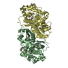
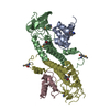
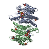
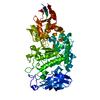
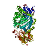
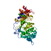
| Title | Crystal structure of mouse pyridoxal kinase in apo form | ||||||
 Components Components | Pyridoxal Kinase | ||||||
 Keywords Keywords | TRANSFERASE / PDXK / PLP / PL / Vitamin B6 / Enzyme | ||||||
| Biological species |  | ||||||
| Method |  X-RAY DIFFRACTION / SYNCHROTRON / MOLECULAR REPLACEMENT / Resolution: 2.45 Å X-RAY DIFFRACTION / SYNCHROTRON / MOLECULAR REPLACEMENT / Resolution: 2.45 Å | ||||||
 Authors Authors | Kasaragod, V.B. / Schindelin, H. | ||||||
| Funding support |  Germany, 1items Germany, 1items
| ||||||
 Citation Citation | Journal: Proc.Natl.Acad.Sci.USA / Year: 2020 Title: Pyridoxal kinase inhibition by artemisinins down-regulates inhibitory neurotransmission. Authors: Kasaragod, V.B. / Pacios-Michelena, A. / Schaefer, N. / Zheng, F. / Bader, N. / Alzheimer, C. / Villmann, C. / Schindelin, H. | ||||||
| History |
|
- Structure visualization
| Structure viewer | Molecule:  MolmilJmol/JSmol MolmilJmol/JSmol |
|---|
- Downloads & links
-Download
| PDBx/mmCIF format | 6yjz.cif.gz | 450.6 KB | Display | PDBx/mmCIF format |
|---|---|---|---|---|
| PDB format | pdb6yjz.ent.gz | 377.2 KB | Display | PDB format |
| PDBx/mmJSON format | 6yjz.json.gz | Tree view | PDBx/mmJSON format | |
| Others |  Other downloads Other downloads |
-Validation report
| Arichive directory | https://data.pdbj.org/pub/pdb/validation_reports/yj/6yjzftp://data.pdbj.org/pub/pdb/validation_reports/yj/6yjz | HTTPS FTP |
|---|
-Related structure data
| Related structure data | 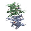 6yk0C 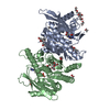 6yk1C  2xytS S: Starting model for refinement C: citing same article ( |
|---|---|
| Similar structure data |


-Links
 PDBj
PDBj- Assembly
| Deposited unit | 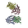
| ||||||||
|---|---|---|---|---|---|---|---|---|---|
| 1 | 
| ||||||||
| 2 | 
| ||||||||
| Unit cell |
|
-Components
| #1: Protein | Mass: 35212.355 Da / Num. of mol.: 4 Source method: isolated from a genetically manipulated source Source: (gene. exp.)  #2: Chemical |   Mass: 194.226 Da / Num. of mol.: 2 / Source method: obtained synthetically / Formula: C8H18O5 / Feature type: SUBJECT OF INVESTIGATION / Comment: precipitant*YM Mass: 194.226 Da / Num. of mol.: 2 / Source method: obtained synthetically / Formula: C8H18O5 / Feature type: SUBJECT OF INVESTIGATION / Comment: precipitant*YM#3: Chemical | ChemComp-GOL /   Mass: 92.094 Da / Num. of mol.: 5 / Source method: obtained synthetically / Formula: C3H8O3 / Feature type: SUBJECT OF INVESTIGATION Mass: 92.094 Da / Num. of mol.: 5 / Source method: obtained synthetically / Formula: C3H8O3 / Feature type: SUBJECT OF INVESTIGATION#4: Chemical | ChemComp-EDO /   Mass: 62.068 Da / Num. of mol.: 15 / Source method: obtained synthetically / Formula: C2H6O2 / Feature type: SUBJECT OF INVESTIGATION Mass: 62.068 Da / Num. of mol.: 15 / Source method: obtained synthetically / Formula: C2H6O2 / Feature type: SUBJECT OF INVESTIGATION#5: Water | ChemComp-HOH / |  Mass: 18.015 Da / Num. of mol.: 187 / Source method: isolated from a natural source / Formula: H2O Mass: 18.015 Da / Num. of mol.: 187 / Source method: isolated from a natural source / Formula: H2OHas ligand of interest | Y | Has protein modification | Y | |
|---|
-Experimental details
-Experiment
| Experiment | Method: X-RAY DIFFRACTION / Number of used crystals: 1 |
|---|
- Sample preparation
| Crystal | Density Matthews: 2.99 Å3/Da / Density % sol: 58.8 % |
|---|---|
| Crystal grow | Temperature: 293 K / Method: vapor diffusion, sitting drop / Details: 0.18-0.24 M sodium thiocyanate and 18-26% PEG 3350 |
-Data collection
| Diffraction | Mean temperature: 100 K / Serial crystal experiment: N |
|---|---|
| Diffraction source | Source: SYNCHROTRON / Site: ESRF  / Beamline: ID23-1 / Wavelength: 0.9724 Å / Beamline: ID23-1 / Wavelength: 0.9724 Å |
| Detector | Type: DECTRIS PILATUS 6M / Detector: PIXEL / Date: Nov 2, 2017 |
| Radiation | Protocol: SINGLE WAVELENGTH / Monochromatic (M) / Laue (L): M / Scattering type: x-ray |
| Radiation wavelength | Wavelength: 0.9724 Å / Relative weight: 1 |
| Reflection | Resolution: 2.45→47.32 Å / Num. obs: 57180 / % possible obs: 95.6 % / Redundancy: 3.2 % / CC1/2: 0.993 / Rmerge(I) obs: 0.098 / Rpim(I) all: 0.093 / Rrim(I) all: 0.08752 / Net I/σ(I): 9.1 |
| Reflection shell | Resolution: 2.45→2.53 Å / Redundancy: 1.9 % / Rmerge(I) obs: 0.75 / Mean I/σ(I) obs: 1.6 / Num. unique obs: 5965 / CC1/2: 0.572 / Rpim(I) all: 0.7 / Rrim(I) all: 0.6776 / % possible all: 99.7 |
- Processing
| Software |
| ||||||||||||||||||||||||||||||||||||||||||||||||||||||||||||||||||||||||||||||||||||||||||||||||||||||||||||||||||||||||||||||||||||||||||||||||||||||||||
|---|---|---|---|---|---|---|---|---|---|---|---|---|---|---|---|---|---|---|---|---|---|---|---|---|---|---|---|---|---|---|---|---|---|---|---|---|---|---|---|---|---|---|---|---|---|---|---|---|---|---|---|---|---|---|---|---|---|---|---|---|---|---|---|---|---|---|---|---|---|---|---|---|---|---|---|---|---|---|---|---|---|---|---|---|---|---|---|---|---|---|---|---|---|---|---|---|---|---|---|---|---|---|---|---|---|---|---|---|---|---|---|---|---|---|---|---|---|---|---|---|---|---|---|---|---|---|---|---|---|---|---|---|---|---|---|---|---|---|---|---|---|---|---|---|---|---|---|---|---|---|---|---|---|---|---|
| Refinement | Method to determine structure: MOLECULAR REPLACEMENT Starting model: 2XYT Resolution: 2.45→47.32 Å / SU ML: 0.33 / Cross valid method: FREE R-VALUE / σ(F): 1.34 / Phase error: 27
| ||||||||||||||||||||||||||||||||||||||||||||||||||||||||||||||||||||||||||||||||||||||||||||||||||||||||||||||||||||||||||||||||||||||||||||||||||||||||||
| Solvent computation | Shrinkage radii: 0.9 Å / VDW probe radii: 1.11 Å | ||||||||||||||||||||||||||||||||||||||||||||||||||||||||||||||||||||||||||||||||||||||||||||||||||||||||||||||||||||||||||||||||||||||||||||||||||||||||||
| Refinement step | Cycle: LAST / Resolution: 2.45→47.32 Å
| ||||||||||||||||||||||||||||||||||||||||||||||||||||||||||||||||||||||||||||||||||||||||||||||||||||||||||||||||||||||||||||||||||||||||||||||||||||||||||
| Refine LS restraints |
| ||||||||||||||||||||||||||||||||||||||||||||||||||||||||||||||||||||||||||||||||||||||||||||||||||||||||||||||||||||||||||||||||||||||||||||||||||||||||||
| LS refinement shell |
|
