Resolution: 2.45→29.9 Å / Num. obs: 25288 / % possible obs: 99.7 % / Redundancy: 3.7 % / Rmerge(I) obs: 0.087 / Rsym value: 0.087 / Net I/σ(I): 6.6
Reflection shell
Diffraction-ID: 1
Resolution (Å)
% possible obs (%)
Redundancy (%)
Rmerge(I) obs
Mean I/σ(I) obs
Num. measured obs
Rsym value
2.45-2.51
100
3.7
0.657
1.1
1858
0.657
2.51-2.58
100
3.7
0.569
1.3
1793
0.569
2.58-2.66
100
3.7
0.485
1.5
1773
0.485
2.66-2.74
100
3.7
0.385
1.8
1686
0.385
2.74-2.83
100
3.7
0.322
2.3
1644
0.322
2.83-2.93
100
3.7
0.249
3
1603
0.249
2.93-3.04
100
3.7
0.181
4.1
1559
0.181
3.04-3.16
100
3.7
0.152
4.8
1488
0.152
3.16-3.3
100
3.7
0.125
5.7
1445
0.125
3.3-3.46
99.9
3.7
0.094
7.4
1361
0.094
3.46-3.65
99.9
3.7
0.08
8.6
1327
0.08
3.65-3.87
99.8
3.7
0.07
8.5
1227
0.07
3.87-4.14
99.7
3.7
0.064
9.1
1156
0.064
4.14-4.47
99.6
3.6
0.057
10.7
1104
0.057
4.47-4.9
99.5
3.6
0.052
11.2
999
0.052
4.9-5.48
99.3
3.7
0.053
9.9
914
0.053
5.48-6.33
99.1
3.6
0.062
10.3
819
0.062
6.33-7.75
98.6
3.5
0.055
11.5
688
0.055
7.75-10.96
98.1
3.5
0.039
15.9
544
0.039
10.96-29.9
90.9
3.2
0.05
11.3
300
0.05
-
Phasing
Phasing
Method: MAD
-
Processing
Software
Name
Version
Classification
NB
REFMAC
5.2.0005
refinement
SCALA
datascaling
PDB_EXTRACT
1.601
dataextraction
MOSFLM
datareduction
CCP4
(SCALA)
datascaling
SOLVE
phasing
Refinement
Method to determine structure: MAD / Resolution: 2.45→29.9 Å / Cor.coef. Fo:Fc: 0.956 / Cor.coef. Fo:Fc free: 0.917 / SU B: 17.201 / SU ML: 0.189 / TLS residual ADP flag: LIKELY RESIDUAL / Cross valid method: THROUGHOUT / ESU R: 0.36 / ESU R Free: 0.267 Stereochemistry target values: MAXIMUM LIKELIHOOD WITH PHASES Details: 1. HYDROGENS HAVE BEEN ADDED IN THE RIDING POSITIONS. 2. A MET-INHIBITION PROTOCOL WAS USED FOR SELENOMETHIONINE INCORPORATION DURING PROTEIN EXPRESSION. THE OCCUPANCY OF THE SE ATOMS IN THE ...Details: 1. HYDROGENS HAVE BEEN ADDED IN THE RIDING POSITIONS. 2. A MET-INHIBITION PROTOCOL WAS USED FOR SELENOMETHIONINE INCORPORATION DURING PROTEIN EXPRESSION. THE OCCUPANCY OF THE SE ATOMS IN THE MSE RESIDUES WAS REDUCED TO 0.7 TO ACCOUNT FOR THE REDUCED SCATTERING POWER DUE TO PARTIAL S-MET INCORPORATION. 3. BOTH PHOSPHATE AND SULFATE IONS ARE PRESENT IN THE CRYSTALLIZATION SOLUTION; SO4 WERE MODELLED INTO DENSITY DUE TO ITS HIGHER CONCENTRATION. IT IS NOT POSSIBLE TO ASSIGNED THE TRUE IDENTITIES OF THESE IONS BASED ON DENSITIY MAP. 4. A43-49, A115-122, A253-257, B43-49, B113-B120 AS WELL AS N-TERMINI ARE NOT PRESENT IN THE MODEL. THERE ARE SOME DISORDERED DENSITIES FOR THE LOOPS BETWEEN A114-123, B110-127 AND A253-257. 5. TWO ATOMS ASSIGNED AS WATERS 98 AND 99 ARE LOCATED IN THE ACTIVE SITES, THE CORRESPONDING ATOMS IN THE STRUCTURAL HOMOLOG 1XFI ARE MAGNESIUM IONS.
Rfactor
Num. reflection
% reflection
Selection details
Rfree
0.255
1288
5.1 %
RANDOM
Rwork
0.189
-
-
-
all
0.192
-
-
-
obs
0.19189
23975
99.43 %
-
Solvent computation
Ion probe radii: 0.8 Å / Shrinkage radii: 0.8 Å / VDW probe radii: 1.2 Å / Solvent model: BABINET MODEL WITH MASK
In the structure databanks used in Yorodumi, some data are registered as the other names, "COVID-19 virus" and "2019-nCoV". Here are the details of the virus and the list of structure data.
Jan 31, 2019. EMDB accession codes are about to change! (news from PDBe EMDB page)
EMDB accession codes are about to change! (news from PDBe EMDB page)
The allocation of 4 digits for EMDB accession codes will soon come to an end. Whilst these codes will remain in use, new EMDB accession codes will include an additional digit and will expand incrementally as the available range of codes is exhausted. The current 4-digit format prefixed with “EMD-” (i.e. EMD-XXXX) will advance to a 5-digit format (i.e. EMD-XXXXX), and so on. It is currently estimated that the 4-digit codes will be depleted around Spring 2019, at which point the 5-digit format will come into force.
The EM Navigator/Yorodumi systems omit the EMD- prefix.
Related info.:Q: What is EMD? / ID/Accession-code notation in Yorodumi/EM Navigator
Yorodumi is a browser for structure data from EMDB, PDB, SASBDB, etc.
This page is also the successor to EM Navigator detail page, and also detail information page/front-end page for Omokage search.
The word "yorodu" (or yorozu) is an old Japanese word meaning "ten thousand". "mi" (miru) is to see.
Related info.:EMDB / PDB / SASBDB / Comparison of 3 databanks / Yorodumi Search / Aug 31, 2016. New EM Navigator & Yorodumi / Yorodumi Papers / Jmol/JSmol / Function and homology information / Changes in new EM Navigator and Yorodumi
 Movie
Movie Controller

 Yorodumi
 Open data
 Basic information
 Components
 Keywords
 Function and homology information
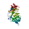
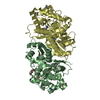
 Archaeoglobus fulgidus (archaea)
 X-RAY DIFFRACTION /
  Authors
 Citation
 Structure visualization
 Downloads & links
 Other downloads


 PDBj
PDBj Assembly


 Mass: 35.453 Da / Num. of mol.: 1 / Source method: obtained synthetically / Formula: Cl
 Mass: 96.063 Da / Num. of mol.: 8 / Source method: obtained synthetically / Formula: SO4
 Mass: 18.015 Da / Num. of mol.: 89 / Source method: isolated from a natural source / Formula: H2O
 Sample preparation
 / Beamline: BL9-2 / Wavelength: 0.97932, 0.97918, 0.91837
 Processing