 Movie
Movie Controller

[English] 日本語
 Yorodumi
- PDB-7a9i: Crystal structure of Coronafacic Acid Ligase from Pectobacterium ... -
+ Open data

- Basic information
| Entry | Database: PDB / ID: 7a9i | ||||||
|---|---|---|---|---|---|---|---|
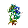
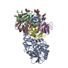
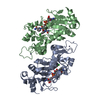
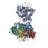
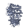
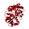
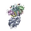
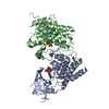
| Title | Crystal structure of Coronafacic Acid Ligase from Pectobacterium brasiliense | ||||||
 Components Components | Cfl | ||||||
 Keywords Keywords | LIGASE | ||||||
| Function / homology |  Function and homology information Function and homology informationmedium-chain fatty acid-CoA ligase activity / fatty acid metabolic process Similarity search - Function | ||||||
| Biological species |  Pectobacterium brasiliense (bacteria) Pectobacterium brasiliense (bacteria) | ||||||
| Method |  X-RAY DIFFRACTION / SYNCHROTRON / MOLECULAR REPLACEMENT / Resolution: 2.1 Å X-RAY DIFFRACTION / SYNCHROTRON / MOLECULAR REPLACEMENT / Resolution: 2.1 Å | ||||||
 Authors Authors | Levy, C.W. | ||||||
| Funding support |  United Kingdom, 1items United Kingdom, 1items
| ||||||
 Citation Citation | Journal: Nature / Year: 2021 Title: Discovery, characterization and engineering of ligases for amide synthesis. Authors: Winn, M. / Rowlinson, M. / Wang, F. / Bering, L. / Francis, D. / Levy, C. / Micklefield, J. #1: Journal: Acta Crystallogr., Sect. D: Biol. Crystallogr. / Year: 2012Title: Towards automated crystallographic structure refinement with phenix.refine. Authors: Afonine, P.V. | ||||||
| History |
|
- Structure visualization
| Structure viewer | Molecule: MolmilJmol/JSmol |
|---|
- Downloads & links
-Download
| PDBx/mmCIF format | 7a9i.cif.gz | 259.3 KB | Display | PDBx/mmCIF format |
|---|---|---|---|---|
| PDB format | pdb7a9i.ent.gz | 173.2 KB | Display | PDB format |
| PDBx/mmJSON format | 7a9i.json.gz | Tree view | PDBx/mmJSON format | |
| Others |  Other downloads Other downloads |
-Validation report
| Arichive directory | https://data.pdbj.org/pub/pdb/validation_reports/a9/7a9iftp://data.pdbj.org/pub/pdb/validation_reports/a9/7a9i | HTTPS FTP |
|---|
-Related structure data


-Links
 PDBj
PDBj
- Assembly
| Deposited unit | 
| ||||||||||||
|---|---|---|---|---|---|---|---|---|---|---|---|---|---|
| 1 |
| ||||||||||||
| Unit cell |
|
-Components
| #1: Protein | Mass: 60406.453 Da / Num. of mol.: 1 Source method: isolated from a genetically manipulated source Source: (gene. exp.) Pectobacterium brasiliense (bacteria) / Gene: cfl, KCO_08370, KU74_07105 / Production host: | ||||
|---|---|---|---|---|---|
| #2: Chemical | ChemComp-R4Z /   Mass: 202.206 Da / Num. of mol.: 1 / Source method: obtained synthetically / Formula: C12H10O3 Mass: 202.206 Da / Num. of mol.: 1 / Source method: obtained synthetically / Formula: C12H10O3 | ||||
| #3: Chemical |   Mass: 94.971 Da / Num. of mol.: 3 / Source method: obtained synthetically / Formula: PO4 Mass: 94.971 Da / Num. of mol.: 3 / Source method: obtained synthetically / Formula: PO4#4: Water | ChemComp-HOH / |  Mass: 18.015 Da / Num. of mol.: 196 / Source method: isolated from a natural source / Formula: H2O Mass: 18.015 Da / Num. of mol.: 196 / Source method: isolated from a natural source / Formula: H2OHas ligand of interest | N | |
-Experimental details
-Experiment
| Experiment | Method: X-RAY DIFFRACTION / Number of used crystals: 1 |
|---|
- Sample preparation
| Crystal | Density Matthews: 2.63 Å3/Da / Density % sol: 53.31 % |
|---|---|
| Crystal grow | Temperature: 277 K / Method: vapor diffusion, sitting drop / pH: 8.5 Details: 2M ammonium sulfate, 0.1M sodium Hepes pH 7.5, 2% v/v Peg 400 Temp details: Cold room |
-Data collection
| Diffraction | Mean temperature: 100 K / Serial crystal experiment: N |
|---|---|
| Diffraction source | Source: SYNCHROTRON / Site: Diamond / Beamline: I04-1 / Wavelength: 0.9159 Å |
| Detector | Type: DECTRIS PILATUS3 6M / Detector: PIXEL / Date: Dec 11, 2018 |
| Radiation | Protocol: SINGLE WAVELENGTH / Monochromatic (M) / Laue (L): M / Scattering type: x-ray |
| Radiation wavelength | Wavelength: 0.9159 Å / Relative weight: 1 |
| Reflection | Resolution: 2.1→64.51 Å / Num. obs: 37973 / % possible obs: 99.82 % / Redundancy: 6.4 % / Biso Wilson estimate: 42.68 Å2 / CC1/2: 0.998 / CC star: 1 / Rmerge(I) obs: 0.1 / Rpim(I) all: 0.043 / Net I/σ(I): 8.07 |
| Reflection shell | Resolution: 2.1→2.175 Å / Redundancy: 6.7 % / Rmerge(I) obs: 1.252 / Mean I/σ(I) obs: 1.4 / Num. unique obs: 3717 / CC1/2: 0.742 / CC star: 0.923 / Rpim(I) all: 0.5237 / % possible all: 99.38 |
- Processing
| Software |
| |||||||||||||||||||||||||||||||||||||||||||||||||||||||||||||||||||||||||||||||||||||||||||||||||||||||||
|---|---|---|---|---|---|---|---|---|---|---|---|---|---|---|---|---|---|---|---|---|---|---|---|---|---|---|---|---|---|---|---|---|---|---|---|---|---|---|---|---|---|---|---|---|---|---|---|---|---|---|---|---|---|---|---|---|---|---|---|---|---|---|---|---|---|---|---|---|---|---|---|---|---|---|---|---|---|---|---|---|---|---|---|---|---|---|---|---|---|---|---|---|---|---|---|---|---|---|---|---|---|---|---|---|---|---|
| Refinement | Method to determine structure: MOLECULAR REPLACEMENT Starting model: inhouse Resolution: 2.1→64.51 Å / SU ML: 0.239 / Cross valid method: FREE R-VALUE / σ(F): 1.33 / Phase error: 26.0074 Stereochemistry target values: GeoStd + Monomer Library + CDL v1.2
| |||||||||||||||||||||||||||||||||||||||||||||||||||||||||||||||||||||||||||||||||||||||||||||||||||||||||
| Solvent computation | Shrinkage radii: 0.9 Å / VDW probe radii: 1.11 Å / Solvent model: FLAT BULK SOLVENT MODEL | |||||||||||||||||||||||||||||||||||||||||||||||||||||||||||||||||||||||||||||||||||||||||||||||||||||||||
| Displacement parameters | Biso mean: 55.88 Å2 | |||||||||||||||||||||||||||||||||||||||||||||||||||||||||||||||||||||||||||||||||||||||||||||||||||||||||
| Refinement step | Cycle: LAST / Resolution: 2.1→64.51 Å
| |||||||||||||||||||||||||||||||||||||||||||||||||||||||||||||||||||||||||||||||||||||||||||||||||||||||||
| Refine LS restraints |
| |||||||||||||||||||||||||||||||||||||||||||||||||||||||||||||||||||||||||||||||||||||||||||||||||||||||||
| LS refinement shell |
| |||||||||||||||||||||||||||||||||||||||||||||||||||||||||||||||||||||||||||||||||||||||||||||||||||||||||
| Refinement TLS params. | Method: refined / Refine-ID: X-RAY DIFFRACTION
| |||||||||||||||||||||||||||||||||||||||||||||||||||||||||||||||||||||||||||||||||||||||||||||||||||||||||
| Refinement TLS group | Refine-ID: X-RAY DIFFRACTION / Auth asym-ID: A
|
