 Movie
Movie Controller


+ Open data

- Basic information
| Entry | Database: PDB / ID: 6u3u | ||||||
|---|---|---|---|---|---|---|---|
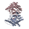
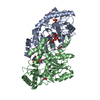
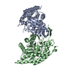
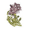
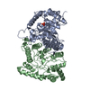
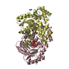
| Title | Crystal Structure of Shiga Toxin 2K | ||||||
 Components Components | (Shiga toxin 2K subunit ...) x 2 | ||||||
 Keywords Keywords | TOXIN / Shiga Toxin / AB5 Toxin / Toxoid | ||||||
| Function / homology |  Function and homology information Function and homology informationsymbiont-mediated hemolysis of host erythrocyte / rRNA N-glycosylase / rRNA N-glycosylase activity / toxin activity / negative regulation of translation / extracellular region / metal ion binding Similarity search - Function | ||||||
| Biological species |  | ||||||
| Method |  X-RAY DIFFRACTION / SYNCHROTRON / MOLECULAR REPLACEMENT / Resolution: 2.287 Å X-RAY DIFFRACTION / SYNCHROTRON / MOLECULAR REPLACEMENT / Resolution: 2.287 Å | ||||||
 Authors Authors | Zhang, Y.Z. / He, X.H. | ||||||
| Funding support |  United States, 1items United States, 1items
| ||||||
 Citation Citation | Journal: Microorganisms / Year: 2019 Title: Structural and Functional Characterization of Stx2k, a New Subtype of Shiga Toxin 2. Authors: Hughes, A.C. / Zhang, Y. / Bai, X. / Xiong, Y. / Wang, Y. / Yang, X. / Xu, Q. / He, X. | ||||||
| History |
|
- Structure visualization
| Structure viewer | Molecule: MolmilJmol/JSmol |
|---|
- Downloads & links
-Download
| PDBx/mmCIF format | 6u3u.cif.gz | 464 KB | Display | PDBx/mmCIF format |
|---|---|---|---|---|
| PDB format | pdb6u3u.ent.gz | 386.1 KB | Display | PDB format |
| PDBx/mmJSON format | 6u3u.json.gz | Tree view | PDBx/mmJSON format | |
| Others |  Other downloads Other downloads |
-Validation report
| Arichive directory | https://data.pdbj.org/pub/pdb/validation_reports/u3/6u3uftp://data.pdbj.org/pub/pdb/validation_reports/u3/6u3u | HTTPS FTP |
|---|
-Related structure data
| Related structure data |  1r4pS S: Starting model for refinement |
|---|---|
| Similar structure data |


-Links
 PDBj
PDBj

- Assembly
| Deposited unit | 
| ||||||||
|---|---|---|---|---|---|---|---|---|---|
| 1 | 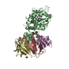
| ||||||||
| 2 | 
| ||||||||
| Unit cell |
|
-Components
-Shiga toxin 2K subunit ... , 2 types, 12 molecules BACDEHIFGJKL
| #1: Protein | Mass: 33038.961 Da / Num. of mol.: 2 / Mutation: E167Q Source method: isolated from a genetically manipulated source Details: E167Q / Source: (gene. exp.) #2: Protein | Mass: 7748.584 Da / Num. of mol.: 10 Source method: isolated from a genetically manipulated source Source: (gene. exp.) Gene: vtx2B, stx2B, stx2B_2, BvCmsKKP036_03423, BvCmsNSP045_04912 Production host: References: UniProt: Q4PS70 |
|---|
-Non-polymers , 4 types, 126 molecules 


| #3: Chemical |  Mass: 238.305 Da / Num. of mol.: 2 / Source method: obtained synthetically / Formula: C8H18N2O4S / Comment: pH buffer*YM Mass: 238.305 Da / Num. of mol.: 2 / Source method: obtained synthetically / Formula: C8H18N2O4S / Comment: pH buffer*YM#4: Chemical | ChemComp-EDO /  Mass: 62.068 Da / Num. of mol.: 7 / Source method: obtained synthetically / Formula: C2H6O2 Mass: 62.068 Da / Num. of mol.: 7 / Source method: obtained synthetically / Formula: C2H6O2#5: Chemical | ChemComp-ZN / |  Mass: 65.409 Da / Num. of mol.: 1 / Source method: obtained synthetically / Formula: Zn Mass: 65.409 Da / Num. of mol.: 1 / Source method: obtained synthetically / Formula: Zn#6: Water | ChemComp-HOH / | Mass: 18.015 Da / Num. of mol.: 116 / Source method: isolated from a natural source / Formula: H2O |
|---|
-Details
| Has ligand of interest | N |
|---|---|
| Has protein modification | Y |
-Experimental details
-Experiment
| Experiment | Method: X-RAY DIFFRACTION / Number of used crystals: 1 |
|---|
- Sample preparation
| Crystal | Density Matthews: 3.35 Å3/Da / Density % sol: 63.26 % |
|---|---|
| Crystal grow | Temperature: 293 K / Method: vapor diffusion, hanging drop / pH: 7.5 Details: 0.1M HEPES, pH 7.5, 10% w/v PEG 8000, 15% Ethylene glycol |
-Data collection
| Diffraction | Mean temperature: 100 K / Serial crystal experiment: N | ||||||||||||||||||||||||||||||
|---|---|---|---|---|---|---|---|---|---|---|---|---|---|---|---|---|---|---|---|---|---|---|---|---|---|---|---|---|---|---|---|
| Diffraction source | Source: SYNCHROTRON / Site: APS / Beamline: 31-ID / Wavelength: 0.9793 Å | ||||||||||||||||||||||||||||||
| Detector | Type: DECTRIS PILATUS 6M / Detector: PIXEL / Date: Feb 13, 2019 | ||||||||||||||||||||||||||||||
| Radiation | Protocol: SINGLE WAVELENGTH / Monochromatic (M) / Laue (L): M / Scattering type: x-ray | ||||||||||||||||||||||||||||||
| Radiation wavelength | Wavelength: 0.9793 Å / Relative weight: 1 | ||||||||||||||||||||||||||||||
| Reflection | Resolution: 2.039→107.059 Å / Num. obs: 94159 / % possible obs: 93.6 % / Redundancy: 3.5 % / CC1/2: 0.998 / Rmerge(I) obs: 0.043 / Rpim(I) all: 0.027 / Rrim(I) all: 0.05 / Net I/σ(I): 13 / Num. measured all: 328715 | ||||||||||||||||||||||||||||||
| Reflection shell | Diffraction-ID: 1
|
- Processing
| Software |
| ||||||||||||||||||||||||||||||||||||||||||||||||||||||||||||||||||||||||||||||||||||||||||
|---|---|---|---|---|---|---|---|---|---|---|---|---|---|---|---|---|---|---|---|---|---|---|---|---|---|---|---|---|---|---|---|---|---|---|---|---|---|---|---|---|---|---|---|---|---|---|---|---|---|---|---|---|---|---|---|---|---|---|---|---|---|---|---|---|---|---|---|---|---|---|---|---|---|---|---|---|---|---|---|---|---|---|---|---|---|---|---|---|---|---|---|
| Refinement | Method to determine structure: MOLECULAR REPLACEMENT Starting model: 1R4P Resolution: 2.287→52.076 Å / SU ML: 0.28 / Cross valid method: THROUGHOUT / σ(F): 1.37 / Phase error: 26.91
| ||||||||||||||||||||||||||||||||||||||||||||||||||||||||||||||||||||||||||||||||||||||||||
| Solvent computation | Shrinkage radii: 0.9 Å / VDW probe radii: 1.11 Å | ||||||||||||||||||||||||||||||||||||||||||||||||||||||||||||||||||||||||||||||||||||||||||
| Displacement parameters | Biso max: 191.64 Å2 / Biso mean: 75.3925 Å2 / Biso min: 39.69 Å2 | ||||||||||||||||||||||||||||||||||||||||||||||||||||||||||||||||||||||||||||||||||||||||||
| Refinement step | Cycle: final / Resolution: 2.287→52.076 Å
| ||||||||||||||||||||||||||||||||||||||||||||||||||||||||||||||||||||||||||||||||||||||||||
| LS refinement shell | Refine-ID: X-RAY DIFFRACTION / Rfactor Rfree error: 0
|
