 Movie
Movie Controller

[English] 日本語
 Yorodumi
- PDB-7mfo: X-ray structure of the L136 Aminotransferase from Acanthamoeba po... -
+ Open data

- Basic information
| Entry | Database: PDB / ID: 7mfo | ||||||
|---|---|---|---|---|---|---|---|
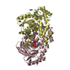
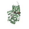
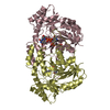
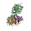
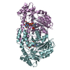
| Title | X-ray structure of the L136 Aminotransferase from Acanthamoeba polyphaga mimivirus in the presence of TDP and PMP | ||||||
 Components Components | L136 aminotransferase | ||||||
 Keywords Keywords | TRANSFERASE / mimivirus / aminotransferase / viosamine | ||||||
| Function / homology |  Function and homology information Function and homology informationpolysaccharide biosynthetic process / transaminase activity / pyridoxal phosphate binding Similarity search - Function | ||||||
| Biological species |   Acanthamoeba polyphaga mimivirus Acanthamoeba polyphaga mimivirus | ||||||
| Method |  X-RAY DIFFRACTION / SYNCHROTRON / SAD / Resolution: 1.7 Å X-RAY DIFFRACTION / SYNCHROTRON / SAD / Resolution: 1.7 Å | ||||||
 Authors Authors | Ferek, J.D. / Thoden, J.B. / Holden, H.M. | ||||||
| Funding support |  United States, 1items United States, 1items
| ||||||
 Citation Citation | Journal: Protein Sci. / Year: 2021 Title: Characterization of an aminotransferase from Acanthamoeba polyphaga Mimivirus. Authors: Seltzner, C.A. / Ferek, J.D. / Thoden, J.B. / Holden, H.M. | ||||||
| History |
|
- Structure visualization
| Structure viewer | Molecule: MolmilJmol/JSmol |
|---|
- Downloads & links
-Download
| PDBx/mmCIF format | 7mfo.cif.gz | 762.5 KB | Display | PDBx/mmCIF format |
|---|---|---|---|---|
| PDB format | pdb7mfo.ent.gz | 627.1 KB | Display | PDB format |
| PDBx/mmJSON format | 7mfo.json.gz | Tree view | PDBx/mmJSON format | |
| Others |  Other downloads Other downloads |
-Validation report
| Arichive directory | https://data.pdbj.org/pub/pdb/validation_reports/mf/7mfoftp://data.pdbj.org/pub/pdb/validation_reports/mf/7mfo | HTTPS FTP |
|---|
-Related structure data


-Links
 PDBj
PDBj- Assembly
| Deposited unit | 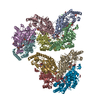
| ||||||||
|---|---|---|---|---|---|---|---|---|---|
| 1 | 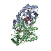
| ||||||||
| 2 | 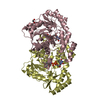
| ||||||||
| 3 | 
| ||||||||
| 4 | 
| ||||||||
| 5 | 
| ||||||||
| Unit cell |
|
-Components
-Protein , 1 types, 10 molecules ABCDEFGHIJ
| #1: Protein | Mass: 40288.754 Da / Num. of mol.: 10 Source method: isolated from a genetically manipulated source Source: (gene. exp.) Acanthamoeba polyphaga mimivirus / Gene: MIMI_L136 / Production host:  |
|---|
-Non-polymers , 6 types, 2944 molecules 


| #2: Chemical | ChemComp-PMP /  Mass: 248.173 Da / Num. of mol.: 10 / Source method: obtained synthetically / Formula: C8H13N2O5P / Feature type: SUBJECT OF INVESTIGATION Mass: 248.173 Da / Num. of mol.: 10 / Source method: obtained synthetically / Formula: C8H13N2O5P / Feature type: SUBJECT OF INVESTIGATION#3: Chemical | ChemComp-PO4 / |  Mass: 94.971 Da / Num. of mol.: 1 / Source method: obtained synthetically / Formula: PO4 Mass: 94.971 Da / Num. of mol.: 1 / Source method: obtained synthetically / Formula: PO4#4: Chemical | ChemComp-EDO /  Mass: 62.068 Da / Num. of mol.: 8 / Source method: obtained synthetically / Formula: C2H6O2 Mass: 62.068 Da / Num. of mol.: 8 / Source method: obtained synthetically / Formula: C2H6O2#5: Chemical | ChemComp-CL / |  Mass: 35.453 Da / Num. of mol.: 1 / Source method: obtained synthetically / Formula: Cl Mass: 35.453 Da / Num. of mol.: 1 / Source method: obtained synthetically / Formula: Cl#6: Chemical | ChemComp-TYD /  Mass: 402.188 Da / Num. of mol.: 10 / Source method: obtained synthetically / Formula: C10H16N2O11P2 Mass: 402.188 Da / Num. of mol.: 10 / Source method: obtained synthetically / Formula: C10H16N2O11P2#7: Water | ChemComp-HOH / | Mass: 18.015 Da / Num. of mol.: 2914 / Source method: isolated from a natural source / Formula: H2O |
|---|
-Details
| Has ligand of interest | Y |
|---|
-Experimental details
-Experiment
| Experiment | Method: X-RAY DIFFRACTION / Number of used crystals: 1 |
|---|
- Sample preparation
| Crystal | Density Matthews: 2.43 Å3/Da / Density % sol: 49.44 % |
|---|---|
| Crystal grow | Temperature: 293 K / Method: microbatch / pH: 7.5 Details: protein incubated with 1 mM PLP and 5 mM TDP-4-aminoquinovose. 11-14% PEG-8000, 200 mM KCl, 100 mM HEPES |
-Data collection
| Diffraction | Mean temperature: 100 K / Serial crystal experiment: N |
|---|---|
| Diffraction source | Source: SYNCHROTRON / Site: APS / Beamline: 19-BM / Wavelength: 0.979 Å |
| Detector | Type: ADSC QUANTUM 210r / Detector: CCD / Date: Jun 23, 2018 |
| Radiation | Protocol: SINGLE WAVELENGTH / Monochromatic (M) / Laue (L): M / Scattering type: x-ray |
| Radiation wavelength | Wavelength: 0.979 Å / Relative weight: 1 |
| Reflection | Resolution: 1.7→50 Å / Num. obs: 397722 / % possible obs: 94.3 % / Observed criterion σ(F): 0 / Observed criterion σ(I): 0 / Redundancy: 4.1 % / Rsym value: 0.055 / Net I/σ(I): 36.2 |
| Reflection shell | Resolution: 1.7→1.73 Å / Redundancy: 2.9 % / Mean I/σ(I) obs: 5 / Num. unique obs: 18360 / Rsym value: 0.194 / % possible all: 87.4 |
- Processing
| Software |
| ||||||||||||||||||||||||||||||||||||||||||||||||||||||||||||
|---|---|---|---|---|---|---|---|---|---|---|---|---|---|---|---|---|---|---|---|---|---|---|---|---|---|---|---|---|---|---|---|---|---|---|---|---|---|---|---|---|---|---|---|---|---|---|---|---|---|---|---|---|---|---|---|---|---|---|---|---|---|
| Refinement | Method to determine structure: SAD / Resolution: 1.7→33.78 Å / Cor.coef. Fo:Fc: 0.957 / Cor.coef. Fo:Fc free: 0.938 / SU B: 2.372 / SU ML: 0.078 / Cross valid method: THROUGHOUT / σ(F): 0 / ESU R: 0.113 / ESU R Free: 0.113 / Stereochemistry target values: MAXIMUM LIKELIHOOD Details: HYDROGENS HAVE BEEN ADDED IN THE RIDING POSITIONS U VALUES : REFINED INDIVIDUALLY
| ||||||||||||||||||||||||||||||||||||||||||||||||||||||||||||
| Solvent computation | Ion probe radii: 0.8 Å / Shrinkage radii: 0.8 Å / VDW probe radii: 1.2 Å / Solvent model: MASK | ||||||||||||||||||||||||||||||||||||||||||||||||||||||||||||
| Displacement parameters | Biso max: 102.23 Å2 / Biso mean: 24.036 Å2 / Biso min: 10.09 Å2
| ||||||||||||||||||||||||||||||||||||||||||||||||||||||||||||
| Refinement step | Cycle: final / Resolution: 1.7→33.78 Å
| ||||||||||||||||||||||||||||||||||||||||||||||||||||||||||||
| Refine LS restraints |
| ||||||||||||||||||||||||||||||||||||||||||||||||||||||||||||
| LS refinement shell | Resolution: 1.7→1.744 Å / Rfactor Rfree error: 0 / Total num. of bins used: 20
|
