| Entry | Database: PDB / ID: 4zas
|
|---|
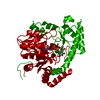
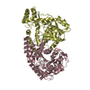
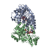
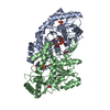
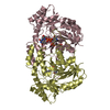
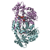
| Title | Crystal structure of sugar aminotransferase CalS13 from Micromonospora echinospora |
|---|
 Components Components | CalS13 |
|---|
 Keywords Keywords | TRANSFERASE / sugar aminotransferase / Structural Genomics / PSI-Biology / Protein Structure Initiative / Enzyme Discovery for Natural Product Biosynthesis / NatPro |
|---|
| Function / homology |  Function and homology information Function and homology information
DegT/DnrJ/EryC1/StrS aminotransferase / DegT/DnrJ/EryC1/StrS aminotransferase family / Aspartate Aminotransferase, domain 1 / Aspartate Aminotransferase, domain 1 / Aspartate Aminotransferase; domain 2 / Type I PLP-dependent aspartate aminotransferase-like (Major domain) / Pyridoxal phosphate-dependent transferase, small domain / Pyridoxal phosphate-dependent transferase, major domain / Pyridoxal phosphate-dependent transferase / Alpha-Beta Complex ...DegT/DnrJ/EryC1/StrS aminotransferase / DegT/DnrJ/EryC1/StrS aminotransferase family / Aspartate Aminotransferase, domain 1 / Aspartate Aminotransferase, domain 1 / Aspartate Aminotransferase; domain 2 / Type I PLP-dependent aspartate aminotransferase-like (Major domain) / Pyridoxal phosphate-dependent transferase, small domain / Pyridoxal phosphate-dependent transferase, major domain / Pyridoxal phosphate-dependent transferase / Alpha-Beta Complex / 3-Layer(aba) Sandwich / Alpha BetaSimilarity search - Domain/homology |
|---|
| Biological species |  Micromonospora echinospora (bacteria) Micromonospora echinospora (bacteria) |
|---|
| Method |  X-RAY DIFFRACTION / SYNCHROTRON / Resolution: 2.47 Å X-RAY DIFFRACTION / SYNCHROTRON / Resolution: 2.47 Å |
|---|
 Authors Authors | Wang, F. / Singh, S. / Miller, M.D. / Thorson, J.S. / Phillips Jr., G.N. / Enzyme Discovery for Natural Product Biosynthesis (NatPro) |
|---|
| Funding support |  United States, 2items United States, 2items | Organization | Grant number | Country |
|---|
| National Institutes of Health/National Institute of General Medical Sciences (NIH/NIGMS) | U01GM098248 | United States | | National Institutes of Health/National Cancer Institute (NIH/NCI) | CA84374 | United States |
|
|---|
 Citation Citation | Journal: To Be Published
Title: Structure characterization of sugar aminotransferases CalS13 and WecE provides the basis for a unifying structural model for stereochemical outcome.
Authors: Wang, F. / Singh, S. / Miller, M.D. / Thorson, J.S. / Phillips Jr., G.N. |
|---|
| History | | Deposition | Apr 13, 2015 | Deposition site: RCSB / Processing site: RCSB |
|---|
| Supersession | Apr 29, 2015 | ID: 4YTJ |
|---|
| Revision 1.0 | Apr 29, 2015 | Provider: repository / Type: Initial release |
|---|
| Revision 2.0 | Sep 6, 2017 | Group: Author supporting evidence / Derived calculations ...Author supporting evidence / Derived calculations / Non-polymer description / Source and taxonomy / Structure summary
Category: entity / entity_src_gen ...entity / entity_src_gen / pdbx_audit_support / pdbx_struct_oper_list
Item: _chem_comp.formula / _chem_comp.name ..._chem_comp.formula / _chem_comp.name / _entity.formula_weight / _entity_src_gen.pdbx_alt_source_flag / _pdbx_audit_support.funding_organization / _pdbx_struct_oper_list.symmetry_operation |
|---|
| Revision 2.1 | Dec 4, 2019 | Group: Author supporting evidence / Category: pdbx_audit_support / Item: _pdbx_audit_support.funding_organization |
|---|
| Revision 2.2 | Apr 2, 2025 | Group: Data collection / Database references / Structure summary
Category: chem_comp_atom / chem_comp_bond ...chem_comp_atom / chem_comp_bond / database_2 / pdbx_entry_details / pdbx_modification_feature
Item: _database_2.pdbx_DOI / _database_2.pdbx_database_accession |
|---|
|
|---|
 Movie
Movie Controller

 Yorodumi
 Open data
 Basic information
 Structure visualization
 Downloads & links
 Other downloads


 PDBj
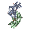
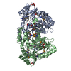
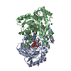
PDBj Assembly
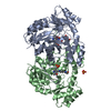

 Mass: 96.063 Da / Num. of mol.: 2 / Source method: obtained synthetically / Formula: SO4
 Mass: 546.314 Da / Num. of mol.: 1 / Source method: obtained synthetically / Formula: C16H24N2O15P2
 Mass: 402.188 Da / Num. of mol.: 3 / Source method: obtained synthetically / Formula: C10H16N2O11P2
 Mass: 18.015 Da / Num. of mol.: 502 / Source method: isolated from a natural source / Formula: H2O
 Sample preparation
 Processing