Mass: 18.015 Da / Num. of mol.: 568 / Source method: isolated from a natural source / Formula: H2O
Has protein modification
Y
Sequence details
THE CONSTRUCT WAS EXPRESSED WITH A PURIFICATION TAG MGSDKIHHHHHHENLYFQG. THE TAG WAS REMOVED WITH ...THE CONSTRUCT WAS EXPRESSED WITH A PURIFICATION TAG MGSDKIHHHHHHENLYFQG. THE TAG WAS REMOVED WITH TEV PROTEASE LEAVING ONLY A GLYCINE (0) FOLLOWED BY THE TARGET SEQUENCE.
-
Experimental details
-
Experiment
Experiment
Method: X-RAY DIFFRACTION / Number of used crystals: 1
-
Sample preparation
Crystal
Density Matthews: 3.39 Å3/Da / Density % sol: 63.74 %
Resolution: 2.6→29.647 Å / Num. obs: 62018 / % possible obs: 97.4 % / Observed criterion σ(I): -3 / Biso Wilson estimate: 59.45 Å2
Reflection shell
Resolution: 2.6→2.69 Å / Rmerge(I) obs: 1.144 / Mean I/σ(I) obs: 1.6 / % possible all: 93.4
-
Phasing
Phasing
Method: MAD
-
Processing
Software
Name
Version
Classification
NB
SHELX
phasing
BUSTER-TNT
BUSTER2.8.0
refinement
XSCALE
datascaling
PDB_EXTRACT
3.1
dataextraction
XDS
datareduction
SHELXD
phasing
autoSHARP
phasing
BUSTER
2.8.0
refinement
Refinement
Method to determine structure: MAD / Resolution: 2.6→29.65 Å / Cor.coef. Fo:Fc: 0.935 / Cor.coef. Fo:Fc free: 0.906 / Occupancy max: 1 / Occupancy min: 0.33 Details: 1. A MET-INHIBITION PROTOCOL WAS USED FOR SELENOMETHIONINE INCORPORATION DURING PROTEIN EXPRESSION. THE OCCUPANCY OF THE SE ATOMS IN THE MSE RESIDUES WAS REDUCED TO 0.75 FOR THE REDUCED ...Details: 1. A MET-INHIBITION PROTOCOL WAS USED FOR SELENOMETHIONINE INCORPORATION DURING PROTEIN EXPRESSION. THE OCCUPANCY OF THE SE ATOMS IN THE MSE RESIDUES WAS REDUCED TO 0.75 FOR THE REDUCED SCATTERING POWER DUE TO PARTIAL S-MET INCORPORATION. 3. ATOM RECORD CONTAINS SUM OF TLS AND RESIDUAL B FACTORS. ANISOU RECORD CONTAINS SUM OF TLS AND RESIDUAL U FACTORS. 4. WATERS WERE EXCLUDED FROM AUTOMATIC TLS ASSIGNMENT. 5. SULFATE (SO4) AND L(+)-TARTARIC ACID (TLA) FROM THE CRYSTALLIZATION SOLUTION AND ETHYLENE GLYCOL USED AS A CRYOPROTECTANT WERE MODELED INTO THE STRUCTURE.
Rfactor
Num. reflection
% reflection
Rfree
0.234
3131
5.05 %
Rwork
0.2
-
-
obs
0.202
61978
-
Displacement parameters
Biso mean: 43.03 Å2
Baniso -1
Baniso -2
Baniso -3
1-
-3.9754 Å2
0 Å2
0 Å2
2-
-
-3.9754 Å2
0 Å2
3-
-
-
7.9507 Å2
Refinement step
Cycle: LAST / Resolution: 2.6→29.65 Å
Protein
Nucleic acid
Ligand
Solvent
Total
Num. atoms
9960
0
121
568
10649
Refine LS restraints
Refine-ID
Type
Number
Restraint function
Weight
Dev ideal
X-RAY DIFFRACTION
t_dihedral_angle_d
4894
SINUSOIDAL
6
X-RAY DIFFRACTION
t_trig_c_planes
284
HARMONIC
2
X-RAY DIFFRACTION
t_gen_planes
1565
HARMONIC
5
X-RAY DIFFRACTION
t_it
10346
HARMONIC
20
X-RAY DIFFRACTION
t_nbd
X-RAY DIFFRACTION
t_improper_torsion
X-RAY DIFFRACTION
t_pseud_angle
X-RAY DIFFRACTION
t_chiral_improper_torsion
1294
SEMIHARMONIC
5
X-RAY DIFFRACTION
t_sum_occupancies
X-RAY DIFFRACTION
t_utility_distance
X-RAY DIFFRACTION
t_utility_angle
X-RAY DIFFRACTION
t_utility_torsion
X-RAY DIFFRACTION
t_ideal_dist_contact
11608
SEMIHARMONIC
4
X-RAY DIFFRACTION
t_bond_d
10346
HARMONIC
2
0.01
X-RAY DIFFRACTION
t_angle_deg
13960
HARMONIC
2
0.98
X-RAY DIFFRACTION
t_omega_torsion
3
X-RAY DIFFRACTION
t_other_torsion
2.48
LS refinement shell
Resolution: 2.6→2.67 Å
Rfactor
Num. reflection
% reflection
Rfree
0.2199
196
4.64 %
Rwork
0.2023
4028
-
all
0.2031
4224
-
Refinement TLS params.
Method: refined / Refine-ID: X-RAY DIFFRACTION
ID
L11 (°2)
L12 (°2)
L13 (°2)
L22 (°2)
L23 (°2)
L33 (°2)
S11 (Å °)
S12 (Å °)
S13 (Å °)
S21 (Å °)
S22 (Å °)
S23 (Å °)
S31 (Å °)
S32 (Å °)
S33 (Å °)
T11 (Å2)
T12 (Å2)
T13 (Å2)
T22 (Å2)
T23 (Å2)
T33 (Å2)
Origin x (Å)
Origin y (Å)
Origin z (Å)
1
0.8845
0.284
-0.1483
0.7992
-0.1605
1.301
0.0219
-0.3075
-0.1698
0.0407
-0.0599
-0.0394
0.1817
-0.0595
0.038
-0.1328
-0.0046
-0.0261
0.1049
0.0766
-0.1655
89.86
-20.3261
34.4252
2
0.9896
-0.139
0.4429
0.3646
0.072
0.5747
0.0347
-0.0402
0.0093
-0.0097
-0.0276
-0.0386
-0.0243
0.0997
-0.007
-0.1141
0.0116
-0.0084
0.1069
0.0063
-0.1061
111.458
0.695
14.8347
3
0.9314
0.1748
0.1469
0.7861
0.1403
1.3061
-0.0348
0.0856
0.0234
-0.1391
0.0163
0.079
-0.0484
-0.258
0.0185
-0.1042
0.0255
-0.0353
0.0141
0.0226
-0.1127
63.6441
6.5095
-38.9527
4
0.3096
0.0208
-0.0688
0.8142
-0.2203
1.1515
-0.0109
-0.0176
0.0357
0.0601
0.0118
-0.0169
-0.3432
0.0506
-0.0009
0.0367
0.0043
-0.0008
-0.0831
0.0065
-0.0869
84.5301
27.4986
-19.3189
Refinement TLS group
ID
Refine-ID
Refine TLS-ID
Selection details
1
X-RAY DIFFRACTION
1
chainA
2
X-RAY DIFFRACTION
2
chainB
3
X-RAY DIFFRACTION
3
chainC
4
X-RAY DIFFRACTION
4
chainD
+
About Yorodumi
-
News
-
Feb 9, 2022. New format data for meta-information of EMDB entries
New format data for meta-information of EMDB entries
Version 3 of the EMDB header file is now the official format.
The previous official version 1.9 will be removed from the archive.
In the structure databanks used in Yorodumi, some data are registered as the other names, "COVID-19 virus" and "2019-nCoV". Here are the details of the virus and the list of structure data.
Jan 31, 2019. EMDB accession codes are about to change! (news from PDBe EMDB page)
EMDB accession codes are about to change! (news from PDBe EMDB page)
The allocation of 4 digits for EMDB accession codes will soon come to an end. Whilst these codes will remain in use, new EMDB accession codes will include an additional digit and will expand incrementally as the available range of codes is exhausted. The current 4-digit format prefixed with “EMD-” (i.e. EMD-XXXX) will advance to a 5-digit format (i.e. EMD-XXXXX), and so on. It is currently estimated that the 4-digit codes will be depleted around Spring 2019, at which point the 5-digit format will come into force.
The EM Navigator/Yorodumi systems omit the EMD- prefix.
Related info.:Q: What is EMD? / ID/Accession-code notation in Yorodumi/EM Navigator
Yorodumi is a browser for structure data from EMDB, PDB, SASBDB, etc.
This page is also the successor to EM Navigator detail page, and also detail information page/front-end page for Omokage search.
The word "yorodu" (or yorozu) is an old Japanese word meaning "ten thousand". "mi" (miru) is to see.
Related info.:EMDB / PDB / SASBDB / Comparison of 3 databanks / Yorodumi Search / Aug 31, 2016. New EM Navigator & Yorodumi / Yorodumi Papers / Jmol/JSmol / Function and homology information / Changes in new EM Navigator and Yorodumi
 Movie
Movie Controller

 Yorodumi
 Open data
 Basic information
 Components
 Keywords
 Function and homology information
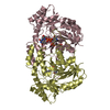
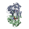
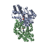
 Burkholderia pseudomallei (bacteria)
 X-RAY DIFFRACTION /
  Authors
 Citation
 Structure visualization
 Downloads & links
 Other downloads


 PDBj
PDBj
 Assembly


 Mass: 96.063 Da / Num. of mol.: 4 / Source method: obtained synthetically / Formula: SO4
 Mass: 150.087 Da / Num. of mol.: 5 / Source method: obtained synthetically / Formula: C4H6O6
 Mass: 62.068 Da / Num. of mol.: 13 / Source method: obtained synthetically / Formula: C2H6O2
 Mass: 18.015 Da / Num. of mol.: 568 / Source method: isolated from a natural source / Formula: H2O
 Sample preparation
 / Beamline: BL9-2 / Wavelength: 0.91837,0.97936,0.97922
 Processing