 Movie
Movie Controller

[English] 日本語
 Yorodumi
- PDB-4piw: Crystal structure of sugar aminotransferase WecE from Escherichia... -
+ Open data

- Basic information
| Entry | Database: PDB / ID: 4piw | |||||||||
|---|---|---|---|---|---|---|---|---|---|---|
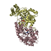
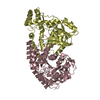
| Title | Crystal structure of sugar aminotransferase WecE from Escherichia coli K-12 | |||||||||
 Components Components | TDP-4-keto-6-deoxy-D-glucose transaminase family protein | |||||||||
 Keywords Keywords | TRANSFERASE / sugar aminotransferase / Structural Genomics / PSI-Biology / Protein Structure Initiative / Enzyme Discovery for Natural Product Biosynthesis / NatPro | |||||||||
| Function / homology |  Function and homology information Function and homology informationdTDP-4-amino-4,6-dideoxygalactose transaminase / dTDP-4-amino-4,6-dideoxygalactose:2-oxoglutarate transaminase activity / enterobacterial common antigen biosynthetic process / polysaccharide biosynthetic process / pyridoxal phosphate binding / identical protein binding Similarity search - Function | |||||||||
| Biological species |  | |||||||||
| Method |  X-RAY DIFFRACTION / SYNCHROTRON / MOLECULAR REPLACEMENT / Resolution: 2.7 Å X-RAY DIFFRACTION / SYNCHROTRON / MOLECULAR REPLACEMENT / Resolution: 2.7 Å | |||||||||
 Authors Authors | Wang, F. / Xu, W. / Helmich, K.E. / Singh, S. / Yennamalli, R.M. / Miller, M.D. / Bingman, C.A. / Thorson, J.S. / Phillips Jr., G.N. / Enzyme Discovery for Natural Product Biosynthesis (NatPro) | |||||||||
| Funding support |  United States, 1items United States, 1items
| |||||||||
 Citation Citation | Journal: To Be Published Title: Crystal structure of sugar aminotransferase WecE from Escherichia coli K-12 Authors: Wang, F. / Xu, W. / Helmich, K.E. / Singh, S. / Yennamalli, R.M. / Bingman, C.A. / Miller, M.D. / Thorson, J.S. / Phillips Jr., G.N. | |||||||||
| History |
|
- Structure visualization
| Structure viewer | Molecule: MolmilJmol/JSmol |
|---|
- Downloads & links
-Download
| PDBx/mmCIF format | 4piw.cif.gz | 564.1 KB | Display | PDBx/mmCIF format |
|---|---|---|---|---|
| PDB format | pdb4piw.ent.gz | 468.7 KB | Display | PDB format |
| PDBx/mmJSON format | 4piw.json.gz | Tree view | PDBx/mmJSON format | |
| Others |  Other downloads Other downloads |
-Validation report
| Arichive directory | https://data.pdbj.org/pub/pdb/validation_reports/pi/4piwftp://data.pdbj.org/pub/pdb/validation_reports/pi/4piw | HTTPS FTP |
|---|
-Related structure data
| Related structure data |  1mdoS  3dr7S  3frkS 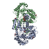 4lc3S 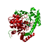 4ocaS S: Starting model for refinement |
|---|---|
| Similar structure data | |
| Other databases |


-Links
 PDBj
PDBj- Assembly
| Deposited unit | 
| ||||||||
|---|---|---|---|---|---|---|---|---|---|
| 1 | 
| ||||||||
| 2 | 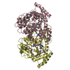
| ||||||||
| 3 | 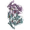
| ||||||||
| 4 | 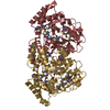
| ||||||||
| Unit cell |
|
-Components
| #1: Protein | Mass: 44955.012 Da / Num. of mol.: 8 Source method: isolated from a genetically manipulated source Source: (gene. exp.) #2: Water | ChemComp-HOH / |  Mass: 18.015 Da / Num. of mol.: 288 / Source method: isolated from a natural source / Formula: H2O Mass: 18.015 Da / Num. of mol.: 288 / Source method: isolated from a natural source / Formula: H2O |
|---|
-Experimental details
-Experiment
| Experiment | Method: X-RAY DIFFRACTION / Number of used crystals: 1 |
|---|
- Sample preparation
| Crystal | Density Matthews: 2.47 Å3/Da / Density % sol: 50.3 % |
|---|---|
| Crystal grow | Temperature: 293 K / Method: vapor diffusion, hanging drop / pH: 6.5 Details: Protein solution (10~25 mg/ml, 25 mM Tris or HEPES pH 7.5, 150 mM NaCl) mixed in a 1:1 ratio with the well solution (0.1 M Sodium Acetate, 0.1 M MES pH6.5, 30% (w/v) PEG 2000 MME), ...Details: Protein solution (10~25 mg/ml, 25 mM Tris or HEPES pH 7.5, 150 mM NaCl) mixed in a 1:1 ratio with the well solution (0.1 M Sodium Acetate, 0.1 M MES pH6.5, 30% (w/v) PEG 2000 MME), cryoprotected with 27% PEG 2000 MME and 10% glycerol |
-Data collection
| Diffraction | Mean temperature: 100 K |
|---|---|
| Diffraction source | Source: SYNCHROTRON / Site: APS / Beamline: 21-ID-F / Wavelength: 0.979 Å |
| Detector | Type: MARMOSAIC 225 mm CCD / Detector: CCD / Date: Apr 17, 2004 |
| Radiation | Monochromator: Diamond [111] / Protocol: SINGLE WAVELENGTH / Monochromatic (M) / Laue (L): M / Scattering type: x-ray |
| Radiation wavelength | Wavelength: 0.979 Å / Relative weight: 1 |
| Reflection twin | Operator: h,-k,-l / Fraction: 0.15 |
| Reflection | Resolution: 2.7→48.8 Å / Num. all: 275071 / Num. obs: 90907 / % possible obs: 84.23 % / Redundancy: 3 % / Rmerge(I) obs: 0.1109 / Net I/σ(I): 10.31 |
- Processing
| Software |
| ||||||||||||||||||||||||||||||||||||||||||||||||||||||||||||||||||||||||||||||||||||
|---|---|---|---|---|---|---|---|---|---|---|---|---|---|---|---|---|---|---|---|---|---|---|---|---|---|---|---|---|---|---|---|---|---|---|---|---|---|---|---|---|---|---|---|---|---|---|---|---|---|---|---|---|---|---|---|---|---|---|---|---|---|---|---|---|---|---|---|---|---|---|---|---|---|---|---|---|---|---|---|---|---|---|---|---|---|
| Refinement | Method to determine structure: MOLECULAR REPLACEMENT Starting model: ensemble model of 1MDO, 3DR7, 3FRK, 4LC3 and 4OCA Resolution: 2.7→48.801 Å / Cross valid method: FREE R-VALUE / σ(F): 0.25 / Phase error: 34.67 / Stereochemistry target values: TWIN_LSQ_F Details: THE ACTIVE SITE CONTENTS ARE LIKELY TO BE A MIXTURE OF NON-BOUND PLP, COVALENTLY BOUND LYSINE-PLP AND PMP. ONLY COVALENTLY BOUND LYSINE-PLP (LLP) WAS BUILT IN DUE TO THE DATA RESOLUTION LIMIT.
| ||||||||||||||||||||||||||||||||||||||||||||||||||||||||||||||||||||||||||||||||||||
| Solvent computation | Shrinkage radii: 0.9 Å / VDW probe radii: 1.11 Å / Solvent model: FLAT BULK SOLVENT MODEL | ||||||||||||||||||||||||||||||||||||||||||||||||||||||||||||||||||||||||||||||||||||
| Displacement parameters | Biso max: 101.93 Å2 / Biso mean: 41.2807 Å2 / Biso min: 5.33 Å2 | ||||||||||||||||||||||||||||||||||||||||||||||||||||||||||||||||||||||||||||||||||||
| Refinement step | Cycle: final / Resolution: 2.7→48.801 Å
| ||||||||||||||||||||||||||||||||||||||||||||||||||||||||||||||||||||||||||||||||||||
| Refine LS restraints |
| ||||||||||||||||||||||||||||||||||||||||||||||||||||||||||||||||||||||||||||||||||||
| LS refinement shell |
|
