 Movie
Movie Controller

[English] 日本語
 Yorodumi
- PDB-6nwr: Thioester acyl-intermediate of Apolipoprotein N-acyltransferase (Lnt) -
+ Open data

- Basic information
| Entry | Database: PDB / ID: 6nwr | ||||||
|---|---|---|---|---|---|---|---|
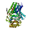
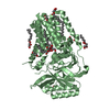
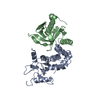
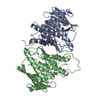
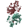
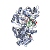
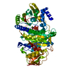
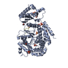
| Title | Thioester acyl-intermediate of Apolipoprotein N-acyltransferase (Lnt) | ||||||
 Components Components | (Apolipoprotein N-acyltransferase) x 2 | ||||||
 Keywords Keywords | MEMBRANE PROTEIN / Enzyme / Nitrilase / N-acyltransferase / Lipoprotein | ||||||
| Function / homology |  Function and homology information Function and homology informationapolipoprotein N-acyltransferase / : / lipoprotein biosynthetic process / outer membrane-bounded periplasmic space / plasma membrane Similarity search - Function | ||||||
| Biological species |  | ||||||
| Method |  X-RAY DIFFRACTION / SYNCHROTRON / MOLECULAR REPLACEMENT / molecular replacement / Resolution: 3.5 Å X-RAY DIFFRACTION / SYNCHROTRON / MOLECULAR REPLACEMENT / molecular replacement / Resolution: 3.5 Å | ||||||
 Authors Authors | Wiseman, B. / Hogbom, M. | ||||||
| Funding support |  Sweden, 1items Sweden, 1items
| ||||||
 Citation Citation | Journal: Sci Rep / Year: 2020 Title: Conformational changes in Apolipoprotein N-acyltransferase (Lnt). Authors: Wiseman, B. / Hogbom, M. | ||||||
| History |
|
- Structure visualization
| Structure viewer | Molecule: MolmilJmol/JSmol |
|---|
- Downloads & links
-Download
| PDBx/mmCIF format | 6nwr.cif.gz | 213.3 KB | Display | PDBx/mmCIF format |
|---|---|---|---|---|
| PDB format | pdb6nwr.ent.gz | 166.5 KB | Display | PDB format |
| PDBx/mmJSON format | 6nwr.json.gz | Tree view | PDBx/mmJSON format | |
| Others |  Other downloads Other downloads |
-Validation report
| Arichive directory | https://data.pdbj.org/pub/pdb/validation_reports/nw/6nwrftp://data.pdbj.org/pub/pdb/validation_reports/nw/6nwr | HTTPS FTP |
|---|
-Related structure data
| Related structure data | 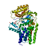 6q3aC  5n6lS C: citing same article ( S: Starting model for refinement |
|---|---|
| Similar structure data |

-Links
 PDBj
PDBj- Assembly
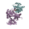
| Deposited unit | 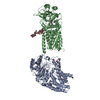
| ||||||||
|---|---|---|---|---|---|---|---|---|---|
| 1 | 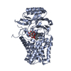
| ||||||||
| 2 | 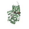
| ||||||||
| Unit cell |
|
-Components
| #1: Protein | Mass: 58988.449 Da / Num. of mol.: 1 Source method: isolated from a genetically manipulated source Source: (gene. exp.) Strain: K12 / Gene: lnt, cutE, b0657, JW0654 / Production host: References: UniProt: P23930, Transferases; Acyltransferases; Transferring groups other than aminoacyl groups | ||
|---|---|---|---|
| #2: Protein | Mass: 59226.855 Da / Num. of mol.: 1 Source method: isolated from a genetically manipulated source Source: (gene. exp.) Strain: K12 / Gene: lnt, cutE, b0657, JW0654 / Production host: References: UniProt: P23930, Transferases; Acyltransferases; Transferring groups other than aminoacyl groups | ||
| #3: Sugar |   Type: D-saccharide / Mass: 510.615 Da / Num. of mol.: 3 / Source method: obtained synthetically / Formula: C24H46O11 / Comment: detergent*YM Type: D-saccharide / Mass: 510.615 Da / Num. of mol.: 3 / Source method: obtained synthetically / Formula: C24H46O11 / Comment: detergent*YM#4: Chemical | ChemComp-D12 / |   Mass: 170.335 Da / Num. of mol.: 1 / Source method: obtained synthetically / Formula: C12H26 Mass: 170.335 Da / Num. of mol.: 1 / Source method: obtained synthetically / Formula: C12H26 |
-Experimental details
-Experiment
| Experiment | Method: X-RAY DIFFRACTION / Number of used crystals: 1 |
|---|
- Sample preparation
| Crystal | Density Matthews: 4.63 Å3/Da / Density % sol: 73.42 % |
|---|---|
| Crystal grow | Temperature: 292 K / Method: vapor diffusion, sitting drop / pH: 6.4 Details: 25 % PEG 2000MME, 100 mM Na cacodylate, 40 mM MgCl2 |
-Data collection
| Diffraction | Mean temperature: 100 K / Serial crystal experiment: N | |||||||||||||||||||||
|---|---|---|---|---|---|---|---|---|---|---|---|---|---|---|---|---|---|---|---|---|---|---|
| Diffraction source | Source: SYNCHROTRON / Site: SLS  / Beamline: X06SA / Wavelength: 1 Å / Beamline: X06SA / Wavelength: 1 Å | |||||||||||||||||||||
| Detector | Type: DECTRIS EIGER X 16M / Detector: PIXEL / Date: Oct 9, 2016 | |||||||||||||||||||||
| Radiation | Protocol: SINGLE WAVELENGTH / Monochromatic (M) / Laue (L): M / Scattering type: x-ray | |||||||||||||||||||||
| Radiation wavelength | Wavelength: 1 Å / Relative weight: 1 | |||||||||||||||||||||
| Reflection | Resolution: 3.5→48.47 Å / Num. obs: 28448 / % possible obs: 99.9 % / Redundancy: 7.4 % / CC1/2: 0.996 / Rmerge(I) obs: 0.277 / Net I/σ(I): 5.1 | |||||||||||||||||||||
| Reflection shell |
|
-Phasing
| Phasing | Method: molecular replacement |
|---|
- Processing
| Software |
| ||||||||||||||||||||||||||||||||||||||||||||||||||||||||||||||||||||||||||||||||||||||||||||||||||||||||||||||||||||||||
|---|---|---|---|---|---|---|---|---|---|---|---|---|---|---|---|---|---|---|---|---|---|---|---|---|---|---|---|---|---|---|---|---|---|---|---|---|---|---|---|---|---|---|---|---|---|---|---|---|---|---|---|---|---|---|---|---|---|---|---|---|---|---|---|---|---|---|---|---|---|---|---|---|---|---|---|---|---|---|---|---|---|---|---|---|---|---|---|---|---|---|---|---|---|---|---|---|---|---|---|---|---|---|---|---|---|---|---|---|---|---|---|---|---|---|---|---|---|---|---|---|---|
| Refinement | Method to determine structure: MOLECULAR REPLACEMENT Starting model: 5N6L Resolution: 3.5→48.469 Å / SU ML: 0.5 / Cross valid method: THROUGHOUT / σ(F): 1.91 / Phase error: 28.2
| ||||||||||||||||||||||||||||||||||||||||||||||||||||||||||||||||||||||||||||||||||||||||||||||||||||||||||||||||||||||||
| Solvent computation | Shrinkage radii: 0.9 Å / VDW probe radii: 1.11 Å | ||||||||||||||||||||||||||||||||||||||||||||||||||||||||||||||||||||||||||||||||||||||||||||||||||||||||||||||||||||||||
| Displacement parameters | Biso max: 215.04 Å2 / Biso mean: 109.6441 Å2 / Biso min: 36.96 Å2 | ||||||||||||||||||||||||||||||||||||||||||||||||||||||||||||||||||||||||||||||||||||||||||||||||||||||||||||||||||||||||
| Refinement step | Cycle: final / Resolution: 3.5→48.469 Å
| ||||||||||||||||||||||||||||||||||||||||||||||||||||||||||||||||||||||||||||||||||||||||||||||||||||||||||||||||||||||||
| LS refinement shell | Refine-ID: X-RAY DIFFRACTION / Rfactor Rfree error: 0 / Total num. of bins used: 19 / % reflection obs: 100 %
|
