 Movie
Movie Controller

[English] 日本語
 Yorodumi
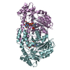
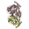
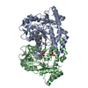
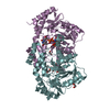
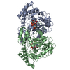
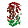
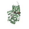
- PDB-1b9h: CRYSTAL STRUCTURE OF 3-AMINO-5-HYDROXYBENZOIC ACID (AHBA) SYNTHASE -
+ Open data

- Basic information
| Entry | Database: PDB / ID: 1b9h | ||||||
|---|---|---|---|---|---|---|---|
| Title | CRYSTAL STRUCTURE OF 3-AMINO-5-HYDROXYBENZOIC ACID (AHBA) SYNTHASE | ||||||
 Components Components | PROTEIN (3-AMINO-5-HYDROXYBENZOIC ACID SYNTHASE) | ||||||
 Keywords Keywords | RIFAMYCIN BIOSYNTHESIS (RIFD GENE) | ||||||
| Function / homology |  Function and homology information Function and homology information3-amino-5-hydroxybenzoate synthase / Transferases; Transferring nitrogenous groups; Transaminases / polysaccharide biosynthetic process / transaminase activity / antibiotic biosynthetic process / pyridoxal phosphate binding / lyase activity Similarity search - Function | ||||||
| Biological species |  Amycolatopsis mediterranei (bacteria) Amycolatopsis mediterranei (bacteria) | ||||||
| Method |  X-RAY DIFFRACTION / SYNCHROTRON / MIR / Resolution: 2 Å X-RAY DIFFRACTION / SYNCHROTRON / MIR / Resolution: 2 Å | ||||||
 Authors Authors | Eads, J.C. / Beeby, M. / Scapin, G. / Yu, T.-W. / Floss, H.G. | ||||||
 Citation Citation | Journal: Biochemistry / Year: 1999 Title: Crystal structure of 3-amino-5-hydroxybenzoic acid (AHBA) synthase. Authors: Eads, J.C. / Beeby, M. / Scapin, G. / Yu, T.W. / Floss, H.G. | ||||||
| History |
|
- Structure visualization
| Structure viewer | Molecule: MolmilJmol/JSmol |
|---|
- Downloads & links
-Download
| PDBx/mmCIF format | 1b9h.cif.gz | 90.2 KB | Display | PDBx/mmCIF format |
|---|---|---|---|---|
| PDB format | pdb1b9h.ent.gz | 68.3 KB | Display | PDB format |
| PDBx/mmJSON format | 1b9h.json.gz | Tree view | PDBx/mmJSON format | |
| Others |  Other downloads Other downloads |
-Validation report
| Arichive directory | https://data.pdbj.org/pub/pdb/validation_reports/b9/1b9hftp://data.pdbj.org/pub/pdb/validation_reports/b9/1b9h | HTTPS FTP |
|---|
-Related structure data


-Links
 PDBj
PDBj- Assembly
| Deposited unit | 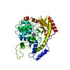
| ||||||||
|---|---|---|---|---|---|---|---|---|---|
| 1 | 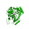
| ||||||||
| Unit cell |
|
-Components
| #1: Protein | Mass: 42262.625 Da / Num. of mol.: 1 Source method: isolated from a genetically manipulated source Source: (gene. exp.) Amycolatopsis mediterranei (bacteria) / Plasmid: PRSETB(EAHBA6) / Production host: |
|---|---|
| #2: Chemical | ChemComp-PLP /   Mass: 247.142 Da / Num. of mol.: 1 / Source method: obtained synthetically / Formula: C8H10NO6P Mass: 247.142 Da / Num. of mol.: 1 / Source method: obtained synthetically / Formula: C8H10NO6P |
| #3: Water | ChemComp-HOH /  Mass: 18.015 Da / Num. of mol.: 216 / Source method: isolated from a natural source / Formula: H2O Mass: 18.015 Da / Num. of mol.: 216 / Source method: isolated from a natural source / Formula: H2O |
-Experimental details
-Experiment
| Experiment | Method: X-RAY DIFFRACTION |
|---|
- Sample preparation
| Crystal | Density Matthews: 3.51 Å3/Da / Density % sol: 60 % Description: MAD DATA USED AS MIR EXPERIMENT, STATISTICS LISTED ARE FOR BEST MAD DATASET (IE NOT NATIVE) | ||||||||||||||||||||||||||||||||||||||||||||||||
|---|---|---|---|---|---|---|---|---|---|---|---|---|---|---|---|---|---|---|---|---|---|---|---|---|---|---|---|---|---|---|---|---|---|---|---|---|---|---|---|---|---|---|---|---|---|---|---|---|---|
| Crystal grow | pH: 7.5 / Details: pH 7.5 | ||||||||||||||||||||||||||||||||||||||||||||||||
| Crystal | *PLUS | ||||||||||||||||||||||||||||||||||||||||||||||||
| Crystal grow | *PLUS Temperature: 18 ℃ / Method: vapor diffusion, hanging drop | ||||||||||||||||||||||||||||||||||||||||||||||||
| Components of the solutions | *PLUS
|
-Data collection
| Diffraction | Mean temperature: 133 K | ||||||||||||
|---|---|---|---|---|---|---|---|---|---|---|---|---|---|
| Diffraction source | Source: SYNCHROTRON / Site: ESRF  / Beamline: BM14 / Wavelength: 1.5418,1.008,0.832 / Beamline: BM14 / Wavelength: 1.5418,1.008,0.832 | ||||||||||||
| Radiation | Protocol: MAD / Monochromatic (M) / Laue (L): M / Scattering type: x-ray | ||||||||||||
| Radiation wavelength |
| ||||||||||||
| Reflection | Resolution: 1.7→99 Å / Num. obs: 65659 / % possible obs: 99.6 % / Redundancy: 1.9 % / Rsym value: 3.8 / Net I/σ(I): 18.6 | ||||||||||||
| Reflection shell | Resolution: 1.7→1.76 Å / Rsym value: 16.6 / % possible all: 100 | ||||||||||||
| Reflection | *PLUS Rmerge(I) obs: 0.037 | ||||||||||||
| Reflection shell | *PLUS % possible obs: 100 % / Rmerge(I) obs: 0.203 / Mean I/σ(I) obs: 2.6 |
- Processing
| Software |
| |||||||||||||||||||||||||||||||||||||||||||||||||||||||||||||||
|---|---|---|---|---|---|---|---|---|---|---|---|---|---|---|---|---|---|---|---|---|---|---|---|---|---|---|---|---|---|---|---|---|---|---|---|---|---|---|---|---|---|---|---|---|---|---|---|---|---|---|---|---|---|---|---|---|---|---|---|---|---|---|---|---|
| Refinement | Method to determine structure: MIR / Resolution: 2→10 Å / Cross valid method: THROUGHOUT / σ(F): 0 Details: XPLOR USED IN EARLIER STAGES OF REFINEMENT. RESIDUE 1 (MET) IS NOT PRESENT IN THE PROTEIN. RESIDUES 2-4 ARE NOT MODELED IN THE STRUCTURE (POOR ELECTRON DENSITY)
| |||||||||||||||||||||||||||||||||||||||||||||||||||||||||||||||
| Refinement step | Cycle: LAST / Resolution: 2→10 Å
| |||||||||||||||||||||||||||||||||||||||||||||||||||||||||||||||
| Refine LS restraints |
| |||||||||||||||||||||||||||||||||||||||||||||||||||||||||||||||
| Software | *PLUS Name: REFMAC / Classification: refinement | |||||||||||||||||||||||||||||||||||||||||||||||||||||||||||||||
| Refinement | *PLUS Highest resolution: 2 Å / σ(F): 0 / % reflection Rfree: 5 % | |||||||||||||||||||||||||||||||||||||||||||||||||||||||||||||||
| Solvent computation | *PLUS | |||||||||||||||||||||||||||||||||||||||||||||||||||||||||||||||
| Displacement parameters | *PLUS |
