 Movie
Movie Controller

[English] 日本語
 Yorodumi
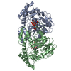
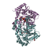
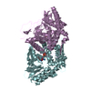
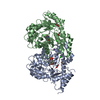
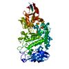
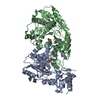
- PDB-3dr4: GDP-perosamine synthase K186A mutant from Caulobacter crescentus ... -
+ Open data

- Basic information
| Entry | Database: PDB / ID: 3dr4 | ||||||
|---|---|---|---|---|---|---|---|
| Title | GDP-perosamine synthase K186A mutant from Caulobacter crescentus with bound sugar ligand | ||||||
 Components Components | Putative perosamine synthetase | ||||||
 Keywords Keywords | TRANSFERASE / Perosamine / deoxysugar / pyridoxal phosphate / aspartate aminotransferase / o-antigen | ||||||
| Function / homology |  Function and homology information Function and homology informationGDP-perosamine synthase / GDP-perosamine synthase activity / O antigen biosynthetic process / pyridoxal phosphate binding Similarity search - Function | ||||||
| Biological species |  Caulobacter crescentus (bacteria) Caulobacter crescentus (bacteria) | ||||||
| Method |  X-RAY DIFFRACTION / SYNCHROTRON / MOLECULAR REPLACEMENT / Resolution: 1.6 Å X-RAY DIFFRACTION / SYNCHROTRON / MOLECULAR REPLACEMENT / Resolution: 1.6 Å | ||||||
 Authors Authors | Holden, H.M. / Cook, P.D. / Carney, A.E. | ||||||
 Citation Citation | Journal: Biochemistry / Year: 2008 Title: Accommodation of GDP-linked sugars in the active site of GDP-perosamine synthase Authors: Cook, P.D. / Carney, A.E. / Holden, H.M. | ||||||
| History |
|
- Structure visualization
| Structure viewer | Molecule: MolmilJmol/JSmol |
|---|
- Downloads & links
-Download
| PDBx/mmCIF format | 3dr4.cif.gz | 326.5 KB | Display | PDBx/mmCIF format |
|---|---|---|---|---|
| PDB format | pdb3dr4.ent.gz | 259.6 KB | Display | PDB format |
| PDBx/mmJSON format | 3dr4.json.gz | Tree view | PDBx/mmJSON format | |
| Others |  Other downloads Other downloads |
-Validation report
| Arichive directory | https://data.pdbj.org/pub/pdb/validation_reports/dr/3dr4ftp://data.pdbj.org/pub/pdb/validation_reports/dr/3dr4 | HTTPS FTP |
|---|
-Related structure data
| Related structure data |  3dr7C  3bn1S S: Starting model for refinement C: citing same article ( |
|---|---|
| Similar structure data |


-Links
 PDBj
PDBj- Assembly
| Deposited unit | 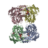
| ||||||||
|---|---|---|---|---|---|---|---|---|---|
| 1 | 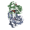
| ||||||||
| 2 | 
| ||||||||
| Unit cell |
|
-Components
| #1: Protein | Mass: 42980.969 Da / Num. of mol.: 4 / Mutation: K186A Source method: isolated from a genetically manipulated source Source: (gene. exp.) Caulobacter crescentus (bacteria) / Strain: CB15 / Gene: per / Plasmid: pET28T / Production host: #2: Chemical | ChemComp-G4M / [( 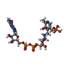  Mass: 819.499 Da / Num. of mol.: 4 / Source method: obtained synthetically / Formula: C24H36N7O19P3 Mass: 819.499 Da / Num. of mol.: 4 / Source method: obtained synthetically / Formula: C24H36N7O19P3#3: Chemical |   Mass: 62.068 Da / Num. of mol.: 2 / Source method: obtained synthetically / Formula: C2H6O2 Mass: 62.068 Da / Num. of mol.: 2 / Source method: obtained synthetically / Formula: C2H6O2#4: Water | ChemComp-HOH / |  Mass: 18.015 Da / Num. of mol.: 1259 / Source method: isolated from a natural source / Formula: H2O Mass: 18.015 Da / Num. of mol.: 1259 / Source method: isolated from a natural source / Formula: H2O |
|---|
-Experimental details
-Experiment
| Experiment | Method: X-RAY DIFFRACTION / Number of used crystals: 1 |
|---|
- Sample preparation
| Crystal | Density Matthews: 2.29 Å3/Da / Density % sol: 46.26 % |
|---|---|
| Crystal grow | Temperature: 298 K / Method: batch / pH: 6.5 Details: 50 mM MES, 10% PEG 8000, 1 mM PLP, 1mM glutamate, pH 6.5, batch, temperature 298K |
-Data collection
| Diffraction | Mean temperature: 100 K |
|---|---|
| Diffraction source | Source: SYNCHROTRON / Site: APS  / Beamline: 19-BM / Wavelength: 0.97 Å / Beamline: 19-BM / Wavelength: 0.97 Å |
| Detector | Type: SBC-3 / Detector: CCD / Date: Feb 8, 2008 |
| Radiation | Monochromator: Si-111 / Protocol: SINGLE WAVELENGTH / Monochromatic (M) / Laue (L): M / Scattering type: x-ray |
| Radiation wavelength | Wavelength: 0.97 Å / Relative weight: 1 |
| Reflection | Resolution: 1.6→50 Å / Num. all: 202983 / Num. obs: 186452 / % possible obs: 91.9 % / Observed criterion σ(F): 2 / Observed criterion σ(I): 2 / Redundancy: 6.3 % / Rsym value: 0.041 / Net I/σ(I): 11.5 |
| Reflection shell | Resolution: 1.6→1.66 Å / Redundancy: 4.6 % / Mean I/σ(I) obs: 7.6 / Num. unique all: 16930 / Rsym value: 0.214 / % possible all: 83.8 |
- Processing
| Software |
| |||||||||||||||||||||||||
|---|---|---|---|---|---|---|---|---|---|---|---|---|---|---|---|---|---|---|---|---|---|---|---|---|---|---|
| Refinement | Method to determine structure: MOLECULAR REPLACEMENT Starting model: PDB entry 3bn1 Resolution: 1.6→26.2 Å / Isotropic thermal model: isotropic / Cross valid method: THROUGHOUT / σ(F): 0 / Stereochemistry target values: Engh & Huber
| |||||||||||||||||||||||||
| Refinement step | Cycle: LAST / Resolution: 1.6→26.2 Å
| |||||||||||||||||||||||||
| Refine LS restraints |
|
