 Movie
Movie Controller


+ Open data

- Basic information
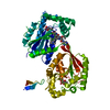
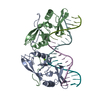
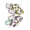
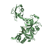
| Entry | Database: PDB / ID: 1ipp | ||||||
|---|---|---|---|---|---|---|---|
| Title | HOMING ENDONUCLEASE/DNA COMPLEX | ||||||
 Components Components |
| ||||||
 Keywords Keywords | TRANSCRIPTION/DNA / HOMING ENDONUCLEASE / INTRON / ZINC / DNA BINDING / PROTEIN FOLDING / TRANSCRIPTION-DNA complex | ||||||
| Function / homology |  Function and homology information Function and homology informationintron homing / endonuclease activity / Hydrolases; Acting on ester bonds / hydrolase activity Similarity search - Function | ||||||
| Biological species |  Physarum polycephalum (eukaryote) Physarum polycephalum (eukaryote) | ||||||
| Method |  X-RAY DIFFRACTION / MIR / Resolution: 2.2 Å X-RAY DIFFRACTION / MIR / Resolution: 2.2 Å | ||||||
 Authors Authors | Flick, K.E. / Jurica, M.S. / Monnat Jr., R.J. / Stoddard, B.L. | ||||||
 Citation Citation | Journal: Nature / Year: 1998 Title: DNA binding and cleavage by the nuclear intron-encoded homing endonuclease I-PpoI. Authors: Flick, K.E. / Jurica, M.S. / Monnat Jr., R.J. / Stoddard, B.L. #1: Journal: Protein Sci. / Year: 1997Title: Crystallization and Preliminary X-Ray Studies of I-Ppoi: A Nuclear, Intron- Encoded Homing Endonuclease from Physarum Polycephalum Authors: Flick, K.E. / Mchugh, D. / Heath, J.D. / Stephens, K.M. / Monnat Jr., R.J. / Stoddard, B.L. | ||||||
| History |
|
- Structure visualization
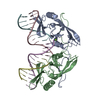
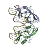
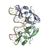
| Structure viewer | Molecule: MolmilJmol/JSmol |
|---|
- Downloads & links
-Download
| PDBx/mmCIF format | 1ipp.cif.gz | 113.6 KB | Display | PDBx/mmCIF format |
|---|---|---|---|---|
| PDB format | pdb1ipp.ent.gz | 82.3 KB | Display | PDB format |
| PDBx/mmJSON format | 1ipp.json.gz | Tree view | PDBx/mmJSON format | |
| Others |  Other downloads Other downloads |
-Validation report
| Arichive directory | https://data.pdbj.org/pub/pdb/validation_reports/ip/1ippftp://data.pdbj.org/pub/pdb/validation_reports/ip/1ipp | HTTPS FTP |
|---|
-Related structure data


-Links
 PDBj
PDBj


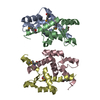
- Assembly
| Deposited unit | 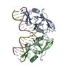
| ||||||||||
|---|---|---|---|---|---|---|---|---|---|---|---|
| 1 |
| ||||||||||
| Unit cell |
| ||||||||||
| Noncrystallographic symmetry (NCS) | NCS oper: (Code: given Matrix: (-0.970715, 0.239393, 0.020068), Vector: |
-Components
| #1: DNA chain | Mass: 6437.184 Da / Num. of mol.: 2 / Source method: obtained synthetically #2: Protein | Mass: 17811.242 Da / Num. of mol.: 2 Source method: isolated from a genetically manipulated source Source: (gene. exp.) Physarum polycephalum (eukaryote) / Cellular location: NUCLEUS / Plasmid: PET / Species (production host): Escherichia coli / Production host:  #3: Chemical | ChemComp-CD /   Mass: 112.411 Da / Num. of mol.: 4 / Source method: obtained synthetically / Formula: Cd Mass: 112.411 Da / Num. of mol.: 4 / Source method: obtained synthetically / Formula: Cd#4: Chemical |   Mass: 24.305 Da / Num. of mol.: 2 / Source method: obtained synthetically / Formula: Mg Mass: 24.305 Da / Num. of mol.: 2 / Source method: obtained synthetically / Formula: Mg#5: Water | ChemComp-HOH / |  Mass: 18.015 Da / Num. of mol.: 547 / Source method: isolated from a natural source / Formula: H2O Mass: 18.015 Da / Num. of mol.: 547 / Source method: isolated from a natural source / Formula: H2OCompound details | DA 213 IN CHAINS C AND D ARE THE SITES OF ENDONUCLEA | |
|---|
-Experimental details
-Experiment
| Experiment | Method: X-RAY DIFFRACTION / Number of used crystals: 1 |
|---|
- Sample preparation
| Crystal | Density Matthews: 3.4 Å3/Da / Density % sol: 60 % | ||||||||||||||||||||||||||||||||||||||||||||||||||||||
|---|---|---|---|---|---|---|---|---|---|---|---|---|---|---|---|---|---|---|---|---|---|---|---|---|---|---|---|---|---|---|---|---|---|---|---|---|---|---|---|---|---|---|---|---|---|---|---|---|---|---|---|---|---|---|---|
| Crystal grow | Method: vapor diffusion / pH: 5.4 Details: THE CRYSTALS WERE GROWN FROM A 2:1 MOLAR RATIO SOLUTION OF PROTEIN AND DNA SUPPLEMENTED WITH 2.5 MM EDTA AND 5 MM SPERMINE. THE COMPLEX WAS CRYSTALLIZED FROM 21 - 27% PEG 4000, 0.1 M ...Details: THE CRYSTALS WERE GROWN FROM A 2:1 MOLAR RATIO SOLUTION OF PROTEIN AND DNA SUPPLEMENTED WITH 2.5 MM EDTA AND 5 MM SPERMINE. THE COMPLEX WAS CRYSTALLIZED FROM 21 - 27% PEG 4000, 0.1 M CITRATE, PH 5.4 - 5.8; 20 MM NACL, 2 MM EDTA., VAPOR DIFFUSION | ||||||||||||||||||||||||||||||||||||||||||||||||||||||
| Components of the solutions |
| ||||||||||||||||||||||||||||||||||||||||||||||||||||||
| Crystal | *PLUS | ||||||||||||||||||||||||||||||||||||||||||||||||||||||
| Crystal grow | *PLUS Temperature: 4 ℃ / Method: vapor diffusion, hanging dropDetails: DNA:protein solution(in 2:1 ratio) was mixed with an equal volume of reservoir solution PH range low: 5.6 / PH range high: 5.4 | ||||||||||||||||||||||||||||||||||||||||||||||||||||||
| Components of the solutions | *PLUS
|
-Data collection
| Diffraction | Mean temperature: 100 K |
|---|---|
| Diffraction source | Source: ROTATING ANODE / Type: RIGAKU RU200 / Wavelength: 1.5418 |
| Detector | Type: RIGAKU RAXIS / Detector: IMAGE PLATE / Date: Aug 15, 1997 / Details: NONE |
| Radiation | Monochromator: NI FILTER / Protocol: SINGLE WAVELENGTH / Monochromatic (M) / Laue (L): M / Scattering type: x-ray |
| Radiation wavelength | Wavelength: 1.5418 Å / Relative weight: 1 |
| Reflection | Resolution: 2.2→50 Å / Num. obs: 30702 / % possible obs: 90 % / Observed criterion σ(I): 2 / Redundancy: 2.7 % / Rmerge(I) obs: 0.048 / Rsym value: 4.8 / Net I/σ(I): 15 |
| Reflection shell | Resolution: 2.2→2.24 Å / Redundancy: 1.3 % / Rmerge(I) obs: 0.193 / Mean I/σ(I) obs: 3.7 / Rsym value: 19.3 / % possible all: 41 |
| Reflection | *PLUS Highest resolution: 2.2 Å / Lowest resolution: 50 Å / % possible obs: 90 % / Observed criterion σ(I): 2 / Redundancy: 2.7 % / Num. measured all: 83203 |
| Reflection shell | *PLUS Highest resolution: 2.2 Å / Lowest resolution: 2.24 Å / Redundancy: 1.3 % / Mean I/σ(I) obs: 3.7 |
- Processing
| Software |
| ||||||||||||||||||||||||||||||||||||||||||||||||||||||||||||
|---|---|---|---|---|---|---|---|---|---|---|---|---|---|---|---|---|---|---|---|---|---|---|---|---|---|---|---|---|---|---|---|---|---|---|---|---|---|---|---|---|---|---|---|---|---|---|---|---|---|---|---|---|---|---|---|---|---|---|---|---|---|
| Refinement | Method to determine structure: MIR / Resolution: 2.2→50 Å / Cross valid method: THROUGHOUT / σ(F): 2
| ||||||||||||||||||||||||||||||||||||||||||||||||||||||||||||
| Displacement parameters | Biso mean: 31.9 Å2 | ||||||||||||||||||||||||||||||||||||||||||||||||||||||||||||
| Refinement step | Cycle: LAST / Resolution: 2.2→50 Å
| ||||||||||||||||||||||||||||||||||||||||||||||||||||||||||||
| Refine LS restraints |
| ||||||||||||||||||||||||||||||||||||||||||||||||||||||||||||
| Refine LS restraints NCS | NCS model details: RESTRAINTS | ||||||||||||||||||||||||||||||||||||||||||||||||||||||||||||
| LS refinement shell | Resolution: 2.2→2.31 Å / Total num. of bins used: 7
| ||||||||||||||||||||||||||||||||||||||||||||||||||||||||||||
| Xplor file |
| ||||||||||||||||||||||||||||||||||||||||||||||||||||||||||||
| Software | *PLUS Name: X-PLOR / Version: 3.8 / Classification: refinement | ||||||||||||||||||||||||||||||||||||||||||||||||||||||||||||
| Refinement | *PLUS Highest resolution: 2.2 Å / Lowest resolution: 50 Å / σ(F): 2 / % reflection Rfree: 5 % / Rfactor obs: 0.212 | ||||||||||||||||||||||||||||||||||||||||||||||||||||||||||||
| Solvent computation | *PLUS | ||||||||||||||||||||||||||||||||||||||||||||||||||||||||||||
| Displacement parameters | *PLUS Biso mean: 31.9 Å2 | ||||||||||||||||||||||||||||||||||||||||||||||||||||||||||||
| Refine LS restraints | *PLUS
| ||||||||||||||||||||||||||||||||||||||||||||||||||||||||||||
| LS refinement shell | *PLUS Highest resolution: 2.2 Å / % reflection Rfree: 4.5 % / Rfactor Rwork: 0.33 |
